Discoveries · Card 20 of 50 · Medicine
Diagnostic Ultrasound: The Glasgow Scanner That Let Doctors See Inside a Living Body
An Austrian neurologist first tried it on the brain in 1942. American engineers built the first workable cross-sectional scanners in the early 1950s. And in a Glasgow maternity ward in 1958, an obstetrician, a 23-year-old engineer and a clinical registrar published the paper — and built the machine — that turned ultrasound from a laboratory curiosity into a routine, life-saving diagnostic tool.
Ian Donald · 1910–1987Tom Brown · engineerUniversity of GlasgowReading time · 20 minUpdated 9 August 2026
In Brief
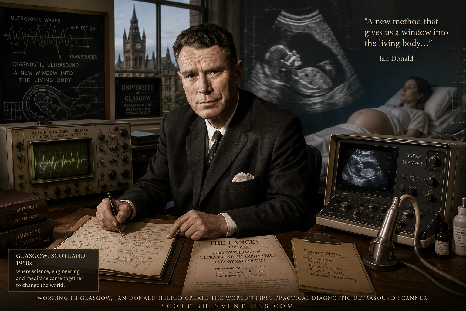
No single country invented diagnostic ultrasound. Karl Theodore Dussik in Austria made the earliest documented attempt to image the living body with ultrasound in 1942, though his transcranial images later proved unreliable. Douglass Howry and Joseph Holmes in the United States built the first working immersion-tank compound scanners in the early 1950s, producing genuinely useful cross-sectional images, while John Wild and John Reid pursued parallel American research into tumour detection with sound waves. What a team at the University of Glasgow — obstetrician Ian Donald, engineer Tom Brown of Kelvin & Hughes, and clinician Dr John MacVicar — achieved between 1955 and 1958 was the first contact compound scanner practical enough for routine hospital use, built specifically for obstetric and abdominal diagnosis, and backed by the systematic clinical evidence published in the 7 June 1958 Lancet paper "Investigation of Abdominal Masses by Pulsed Ultrasound." Glasgow did not invent ultrasound. It invented the first ultrasound scanner and protocol that a working hospital could actually use, day after day, on real patients.
Scottish Impact Index (SI Score)
- Global Impact
- 10/10
- Longevity
- 9/10
- Economic / Material Impact
- 8/10
- Originality / Distinctiveness
- 9/10
- Legacy (Built Upon)
- 9/10
- Recognition
- 6/10
Key Facts
- Discovery
- The first practical clinical contact compound scanner for obstetric and abdominal diagnosis, and the 1958 Lancet paper that founded the field of diagnostic medical ultrasound
- Key figures
- Ian Donald (1910–1987), obstetrician; Tom Brown, engineer, Kelvin & Hughes; Dr John MacVicar, clinical registrar
- Institution
- University of Glasgow / Western Infirmary, with engineering at Kelvin & Hughes, Glasgow
- Landmark publication
- Donald, MacVicar & Brown, 'Investigation of Abdominal Masses by Pulsed Ultrasound,' The Lancet, 7 June 1958
- Key device
- A 'bed-table' contact compound scanner built from an industrial flaw-detector, a hospital bed and Meccano chains; later commercialised as the Diasonograph
- Rival claim — first use of ultrasound in medicine
- Karl Theodore Dussik (Austria), transcranial 'hyperphonography' to image brain ventricles, 1942
- Rival claim — B-mode compound scanning
- Douglass Howry and Joseph Holmes (USA), immersion tank compound scanners, early-to-mid 1950s
- Rival claim — Doppler and pulse-echo research
- John Wild and John Reid (USA/UK-trained), early A-mode and B-mode tumour-detection work, USA, from 1950
- Global impact today
- Ultrasound is among the most widely performed diagnostic imaging examinations in the world, used in obstetrics, cardiology, emergency medicine and beyond
- Claim status
- ESTABLISHED, with a defined scope — Glasgow did not invent ultrasound or medical sonography; it produced the first scanner and protocol practical enough for routine clinical obstetric use, and the foundational 1958 publication
Before Glasgow: Dussik, Howry and Wild
The idea that sound waves could be used to see inside the human body did not begin in Scotland. It began, at least in documented medical use, with the Austrian neurologist and psychiatrist Karl Theodore Dussik, who in 1942 attempted to image the fluid-filled ventricles of the brain by passing ultrasound transcranially and recording the transmitted signal — a technique he called "hyperphonography." It was an extraordinary idea for its time, but later analysis showed that much of what Dussik's images actually captured was the varying thickness of the skull bone rather than genuine detail from inside the brain. The method was never adopted clinically, but the attempt itself earns Dussik a permanent place as the person who first tried to turn ultrasound into a diagnostic tool.
A decade later, in Denver, Colorado, the American researchers Douglass Howry and Joseph Holmes built a genuinely working compound B-mode scanner, in which a patient was partly immersed in a water tank so that ultrasound could be coupled efficiently into the body from multiple angles as a transducer rotated around them. Their apparatus — early versions were built using a modified war-surplus gun turret from a B-29 bomber — produced recognisable cross-sectional images of soft tissue, a real scientific milestone. It was, however, cumbersome, slow, and entirely impractical as a piece of everyday hospital equipment: no maternity ward was going to install an immersion tank and rotating gun turret to check a pregnant patient.
At around the same time, John Wild and John Reid, working in the United States, pursued a parallel line of research using pulse-echo instruments to detect and characterise breast tumours, contributing important early evidence that ultrasound echoes carried genuine diagnostic information about tissue type. None of this American and Austrian work fed directly into the Glasgow project — it developed independently, on the other side of the Atlantic, and this page names it explicitly rather than allowing a Scottish narrative to crowd it out.
Ian Donald's Insight
Ian Donald was born not in Scotland but in Liskeard, Cornwall, on 27 December 1910, to a Scottish medical family with roots in Paisley. Educated partly at Fettes College in Edinburgh and at St Thomas's Hospital Medical School in London, he served as a Royal Air Force medical officer during the Second World War, an experience that left him with a working knowledge of radar and sonar. In 1954 he was appointed Regius Professor of Midwifery at the University of Glasgow, a Crown appointment he took on condition that a new maternity hospital would be built.
In 1950s obstetric practice, distinguishing a benign fluid-filled cyst from a solid, potentially malignant tumour, or judging the position of the placenta, depended largely on a doctor's palpating hands and clinical judgement. Donald's insight was to look outside medicine altogether: ultrasonic flaw-detectors were already used in Clyde shipyards and boiler works to find hidden cracks in welded steel. If sound echoes could reveal a flaw in metal, he reasoned, they might reveal the difference between a cyst and a tumour in flesh. An opportunity arrived through one of his patients, whose husband was a director at the boiler-makers Babcock & Wilcox in Renfrew, who invited Donald to try the firm's industrial equipment.
Tom Brown: The Under-Credited Engineer
If Donald supplied the clinical vision, it was Tom Brown who supplied the engineering that made it real — and Brown's contribution has historically been the most under-credited part of this story. Brown was a 23-year-old research engineer at Kelvin & Hughes, the Glasgow scientific-instruments firm, when he heard in 1956 that an obstetrician was experimenting with one of the company's industrial flaw-detectors, and sought Donald out. Over the following two years Brown designed and built the world's first practical two-dimensional contact compound scanner: a working instrument that could be pressed directly against a patient's skin, rather than requiring immersion in a water tank as Howry and Holmes's American apparatus did.
The device that emerged was, by any glamorous standard, a piece of improvised engineering: a modified Kelvin & Hughes flaw-detector, a cathode-ray display for viewing the returning echoes, a redundant hospital bed repurposed as a mounting frame, and Meccano chains and sprockets used to move the transducer smoothly across the patient. Brown was formally named as inventor on the resulting Kelvin & Hughes patent — a fact that is often left out of accounts that credit the invention of ultrasound to Donald alone. Popular retellings tend to compress a genuine three-person, two-institution collaboration into the story of a single visionary doctor; this page treats Brown as a full co-inventor because the documentary record — the patent, the joint Lancet authorship, and Brown and Donald's joint honorary membership of the British Medical Ultrasound Society in 1984 — supports nothing less.
John MacVicar's Clinical Eye
Dr John MacVicar was Donald's clinical registrar and joined the project in 1956, contributing to scanning sessions, patient assessment, and to the 1958 Lancet paper as its third named author. His clinical judgement mattered in a very concrete way: when a woman referred to Glasgow with a diagnosis of inoperable terminal cancer elsewhere was scanned by the team, MacVicar's reading of the ultrasound trace identified the mass as a benign ovarian cyst rather than a malignant tumour. Surgery based on that finding saved her life, and the case did more to convince sceptical colleagues of ultrasound's value than any amount of theoretical argument could have done.
The Steak Experiment, 1955
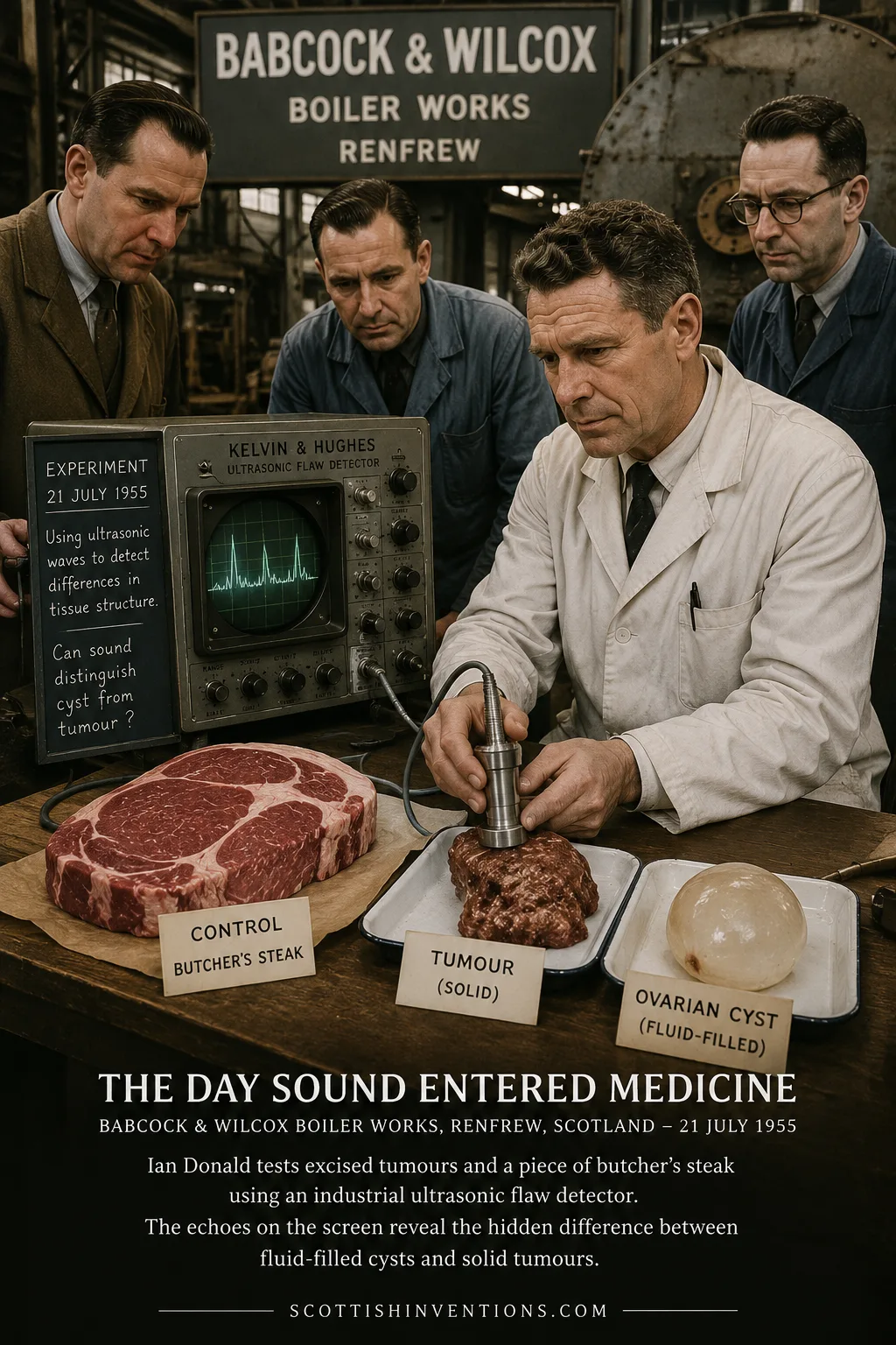
On 21 July 1955, Donald travelled to the Babcock & Wilcox works at Renfrew carrying fibroids and a large ovarian cyst excised from patients that same morning. A technician demonstrated the firm's Kelvin & Hughes flaw-detector by bouncing its ultrasonic beam off the bone of his own thumb. Donald then tested his specimens — and, in the detail that has become part of the story's folklore, a piece of ordinary butcher's steak supplied as a control tissue. The results, he later wrote, were "beyond my wildest dreams": the fluid-filled cyst returned echoes only from its near and far walls, while the solid tumour attenuated the beam progressively with depth. Sound could tell the difference. That single afternoon supplied the proof of principle on which the rest of the project was built.
Building the Contact Scanner
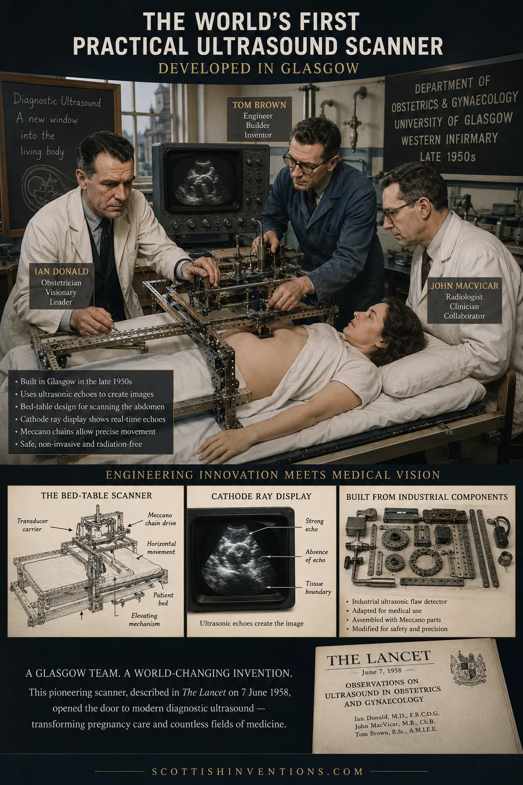
With Brown's engineering and MacVicar's clinical input, the team spent 1956 and 1957 refining a "bed-table" contact compound scanner capable of building up a two-dimensional cross-sectional image by sweeping a transducer smoothly across a patient's abdomen while it stayed in contact with the skin — a deliberate contrast with the American immersion-tank approach. By 1957 the machine was good enough for routine use on real patients at the Western Infirmary, and the case involving the misdiagnosed cyst (described above) demonstrated its clinical value beyond doubt.
A commercial descendant of the Glasgow prototype, refined with the help of industrial designer Dugald Cameron of the Glasgow School of Art, was built at Kelvin & Hughes' Hillington works around 1963–64 and named the Diasonograph. At roughly a ton in weight, it earned hospital staff's affectionate nickname, the "Dinosaurograph" — a reminder of how far the technology still had to travel before it became the small, portable devices used at a patient's bedside today.
The 1958 Lancet Paper
"Investigation of Abdominal Masses by Pulsed Ultrasound" — Ian Donald, J. MacVicar and T. G. Brown. The Lancet, 7 June 1958, vol. 271, no. 7032, pp. 1188–1195.
Across more than nine pages the paper reported findings from around 100 patients and included twelve B-mode illustrations of the gravid uterus, ovarian cysts, fibroids and ascites — among them the first published ultrasound image of a foetus. It has been called, by historians of the field, probably the single most important paper ever published on medical diagnostic ultrasound, not because it revealed a new physical principle but because it supplied the systematic clinical proof — patient after patient — that ultrasound could be trusted to distinguish conditions that genuinely mattered for treatment.
How Ultrasound Works
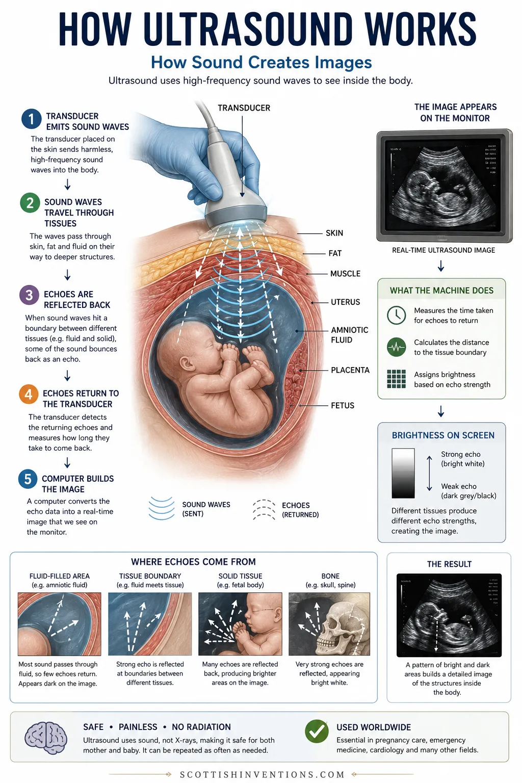
A transducer pressed against the skin emits high-frequency sound pulses, far above the range of human hearing. Whenever those pulses cross a boundary between two different kinds of tissue — fluid and solid, for example, or muscle and bone — some of the sound reflects back toward the transducer as an echo. By timing exactly how long each echo takes to return, and how strong it is, the machine can calculate how far away the boundary lies and how solid or fluid it is, then assign each point a brightness value to build a real-time cross-sectional image. Because it uses ordinary sound waves rather than ionising radiation, ultrasound can be repeated as often as clinically necessary, which is one of the central reasons it became the default imaging method throughout pregnancy.
The Diasonograph and the Spread of the Technology
The Diasonograph, and the Queen Mother's Hospital that opened at Yorkhill, Glasgow in 1964, together made Glasgow an international pilgrimage site for obstetric ultrasound research through the 1960s and 1970s. Visiting clinicians and engineers from around the world came to see the Glasgow technique in action, and manufacturers elsewhere — in the United States, Japan and continental Europe — developed their own commercial scanners, often drawing on the same underlying compound B-mode principles that Glasgow, Howry and Holmes had each separately advanced. It is that combination of an obstetric specialism, a clinically proven protocol and a specific commercial device, rather than a monopoly on the underlying physics, that constitutes Glasgow's lasting contribution.
Legacy: A Billion Scans a Year
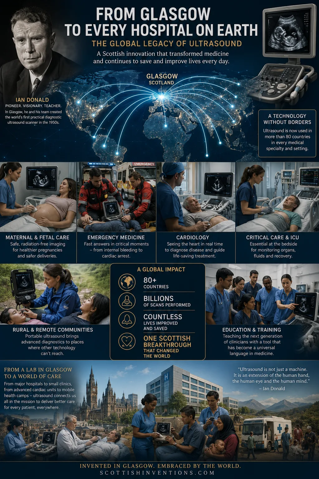
Diagnostic ultrasound is today one of the most frequently performed medical imaging examinations in the world, used far beyond its original obstetric application — in cardiology, emergency medicine, intensive care, musculoskeletal diagnosis and much more. Every pregnant woman who has ever had a scan to check her baby's growth, and every patient who has had a suspicious lump investigated without surgery, has benefited, directly or indirectly, from the clinical protocol and scanner design that Donald, Brown and MacVicar proved could work in a Glasgow hospital in the late 1950s, building on ideas first tested by Dussik in Austria and Howry, Holmes, Wild and Reid in the United States.
Myth vs Evidence
- Myth: "Ultrasound was invented in Scotland." Evidence: No — the underlying physics and the earliest medical attempts to use it belong to Dussik (Austria, 1942) and Howry and Holmes (USA, early 1950s). Glasgow built the first practical clinical contact scanner and supplied the decisive clinical evidence in 1958.
- Myth: "Ian Donald invented ultrasound single-handedly." Evidence: No — Tom Brown was named as inventor on the resulting patent for the scanner itself, and John MacVicar's clinical judgement and co-authorship were essential to the project's success and its 1958 publication.
- Myth: "Ian Donald was Scottish by birth." Evidence: No — he was born in Liskeard, Cornwall, in 1910, though his family and career were deeply rooted in Scotland.
- Myth: "The Glasgow scanner was the first machine ever to attempt medical ultrasound imaging." Evidence: No — Dussik's transcranial experiments in Austria predate it by thirteen years, and Howry and Holmes's American immersion-tank scanners by several years.
Timeline
| Date | Event | Why it matters |
|---|---|---|
| 1942 | Karl Theodore Dussik in Austria attempts transcranial ultrasound imaging of the brain's ventricles | The earliest known attempt to use ultrasound for medical diagnosis, though the images proved unreliable and the technique was not adopted clinically |
| Early 1950s | Douglass Howry and Joseph Holmes in Colorado, USA, build immersion-tank compound B-mode scanners | Produce recognisable cross-sectional images of soft tissue, but the apparatus — including a modified war-surplus gun turret — is bulky and impractical for routine hospital use |
| 1950s | John Wild and John Reid in the USA develop pulse-echo instruments to detect breast tumours and study tissue | Important early work on using ultrasound to distinguish tissue types, running in parallel with Glasgow rather than feeding directly into it |
| 21 July 1955 | Ian Donald tests excised tumours, an ovarian cyst and a butcher's steak against a Kelvin & Hughes industrial flaw-detector at Babcock & Wilcox, Renfrew | Confirms that ultrasound echoes can distinguish fluid-filled cysts from solid tumours in human tissue |
| 1956 | Engineer Tom Brown of Kelvin & Hughes joins Donald; Dr John MacVicar joins as clinical registrar | The three-person Glasgow team is complete; Brown begins engineering a purpose-built two-dimensional contact scanner |
| 1957 | A patient diagnosed elsewhere as having inoperable terminal cancer is scanned in Glasgow | The scan reveals a benign ovarian cyst rather than a tumour; surgery saves her life and wins over sceptical colleagues |
| 7 June 1958 | Donald, MacVicar and Brown publish 'Investigation of Abdominal Masses by Pulsed Ultrasound' in The Lancet | Reports on 100 patients and includes the first published ultrasound image of a foetus; widely regarded as the founding paper of diagnostic medical ultrasound |
| 1963–64 | The Diasonograph, the first commercial descendant of the Glasgow scanner, is built at Kelvin & Hughes' Hillington works | At roughly a ton in weight, hospital staff nickname it the 'Dinosaurograph' |
| 1964 | The Queen Mother's Hospital opens at Yorkhill, Glasgow, campaigned for by Donald | Becomes an international centre for obstetric ultrasound research and training |
| 1984 | Donald and Brown are jointly made the first Honorary Members of the British Medical Ultrasound Society | A rare instance of the engineer receiving formal recognition alongside the clinician |
| Today | Ultrasound is one of the most frequently performed diagnostic imaging examinations worldwide | Used routinely in obstetrics, cardiology, emergency and intensive care, far beyond its original obstetric application |
Frequently Asked Questions
Who invented ultrasound?
No single person or country invented ultrasound imaging, and this site does not claim otherwise. The Austrian neurologist Karl Theodore Dussik made the earliest documented attempt to image the body with ultrasound in 1942, trying to visualise the brain's ventricles, though the results proved unreliable. In the early-to-mid 1950s, Douglass Howry and Joseph Holmes in the United States built immersion-tank compound scanners that produced recognisable cross-sectional images, while John Wild and John Reid separately pursued pulse-echo tumour detection. What a Glasgow team — obstetrician Ian Donald, engineer Tom Brown and clinician John MacVicar — achieved between 1955 and 1958 was the first contact compound scanner practical enough for routine clinical use, applied specifically and successfully to obstetric and abdominal diagnosis, and published in the field-defining 1958 Lancet paper.
Did Scotland invent ultrasound?
Not as such. Scotland did not discover the physical principle that sound waves reflect off tissue boundaries, did not build the first machine to attempt medical ultrasound imaging (that was Dussik, in Austria, in 1942), and did not build the first working compound scanner to produce cross-sectional body images (that was Howry and Holmes, in the United States, in the early 1950s). What the Glasgow team did originate is the first practical, clinically usable contact scanner, built specifically for obstetric and abdominal diagnosis, together with the systematic clinical evidence — a hundred patients, published in The Lancet in 1958 — that made hospitals worldwide take diagnostic ultrasound seriously.
Who was Tom Brown, and why does his role matter so much?
Tom Brown was a 23-year-old research engineer at the Glasgow scientific-instruments firm Kelvin & Hughes when he joined Ian Donald's project in 1956. He designed and built the two-dimensional contact compound scanner that made routine clinical scanning practical, adapting an industrial ultrasonic flaw-detector, a cathode-ray display, a redundant hospital bed and Meccano chains and sprockets into a working diagnostic instrument. Brown was named as inventor on the resulting Kelvin & Hughes patent. Because Donald was the senior clinician and the more prominent public figure, popular accounts have often reduced the story to 'Ian Donald invented ultrasound,' understating that the engineering breakthrough that turned Donald's idea into a usable machine was Brown's. This page treats Brown as a co-inventor, not a footnote.
What was Dr John MacVicar's contribution?
John MacVicar was Donald's clinical registrar in Glasgow. He joined the project in 1956, took part in scanning patients, and was a co-author of the 1958 Lancet paper alongside Donald and Brown. His clinical judgement was decisive in at least one documented case: when a patient referred elsewhere with a diagnosis of inoperable terminal cancer was scanned in Glasgow, MacVicar's assessment of the resulting trace helped identify the mass as a benign ovarian cyst rather than a tumour, and surgery based on that finding saved the patient's life. That case did more than any amount of theory to convince sceptical colleagues that the new technique had real diagnostic value.
What exactly did the 1958 Lancet paper show, and why is it considered so important?
'Investigation of Abdominal Masses by Pulsed Ultrasound,' published by Donald, MacVicar and Brown in The Lancet on 7 June 1958, reported findings from around 100 patients and included twelve illustrations of B-mode ultrasound images of the pregnant uterus, ovarian cysts, fibroids and ascites — among them the first published ultrasound image of a human foetus. It has been described by historians of the field as probably the single most important paper in the history of medical diagnostic ultrasound, not because it introduced a wholly new physical principle, but because it demonstrated, with real clinical patients and a working instrument, that ultrasound imaging could reliably distinguish between conditions that mattered to patient care.
What did earlier researchers such as Dussik, Howry and Wild actually achieve, and why isn't their work usually called 'the invention of ultrasound'?
Karl Theodore Dussik's 1942 transcranial experiments were the earliest recorded attempt to use ultrasound to visualise structures inside the living body, but the resulting images of brain ventricles were later shown to be artefacts of skull bone rather than genuine anatomical detail, and the method was never adopted clinically. Douglass Howry and Joseph Holmes, working independently in Colorado in the early 1950s, built immersion-tank compound scanners — including a famous apparatus improvised from a modified gun turret — that produced genuinely useful cross-sectional images, an important scientific advance, but their equipment was too bulky and slow for routine hospital use. John Wild and John Reid pursued parallel pulse-echo work on tumour detection in the United States in the same period. All of this research is real, important, and predates or runs parallel to Glasgow's work; it is presented here in full rather than omitted, because a Scottish claim to 'first practical clinical obstetric ultrasound' is accurate and defensible, while a claim to have single-handedly invented ultrasound imaging is not.
Was Ian Donald born in Scotland?
No. Despite a persistent myth, Ian Donald was born in Liskeard, Cornwall, on 27 December 1910, to a Scottish medical family with roots in Paisley. He was educated partly at Fettes College in Edinburgh and took his medical degree at St Thomas's Hospital Medical School in London. His entire pioneering work on diagnostic ultrasound, however, took place in Glasgow, where he was appointed Regius Professor of Midwifery at the University of Glasgow in 1954.
Is it fair to call diagnostic ultrasound a 'Scottish invention'?
Only with precision, which is why this page states the scope of the claim plainly rather than leaving it implied. The physical basis of medical sonography and its earliest experimental applications are not Scottish: Dussik's initial 1942 attempt was Austrian, and the immersion-tank compound scanning that first produced usable cross-sectional images was American, built by Howry and Holmes in the early 1950s. What is specifically and defensibly Scottish is the first contact compound scanner practical enough for routine clinical obstetric and abdominal use, engineered by Tom Brown at Kelvin & Hughes to Ian Donald's clinical brief with John MacVicar's diagnostic input, and the 1958 Lancet paper that turned ultrasound from a laboratory curiosity into a technique hospitals adopted. This page presents that narrower, evidence-based claim rather than an unqualified assertion that ultrasound itself was invented in Scotland.
Sources
Tier 1 · Primary
- Donald, I., MacVicar, J. & Brown, T.G., "Investigation of Abdominal Masses by Pulsed Ultrasound," The Lancet, 7 June 1958, vol. 271, no. 7032, pp. 1188–1195.
- Kelvin & Hughes patent records naming Tom Brown as inventor of the contact compound scanner.
Tier 2 · Scholarly and institutional
- Royal College of Physicians and University of Glasgow biographical records on Ian Donald.
- British Medical Ultrasound Society, historical accounts of Donald, Brown and MacVicar.
- Woo, J., "A short History of the development of Ultrasound in Obstetrics and Gynecology," ob-ultrasound.net — covering Dussik, Howry, Holmes, Wild and Reid.