Discoveries · No. 46 of 50 · Medicine
The MRI Scanner: Aberdeen's Spin-Warp Breakthrough — and the Nobel Prize That Left It Out
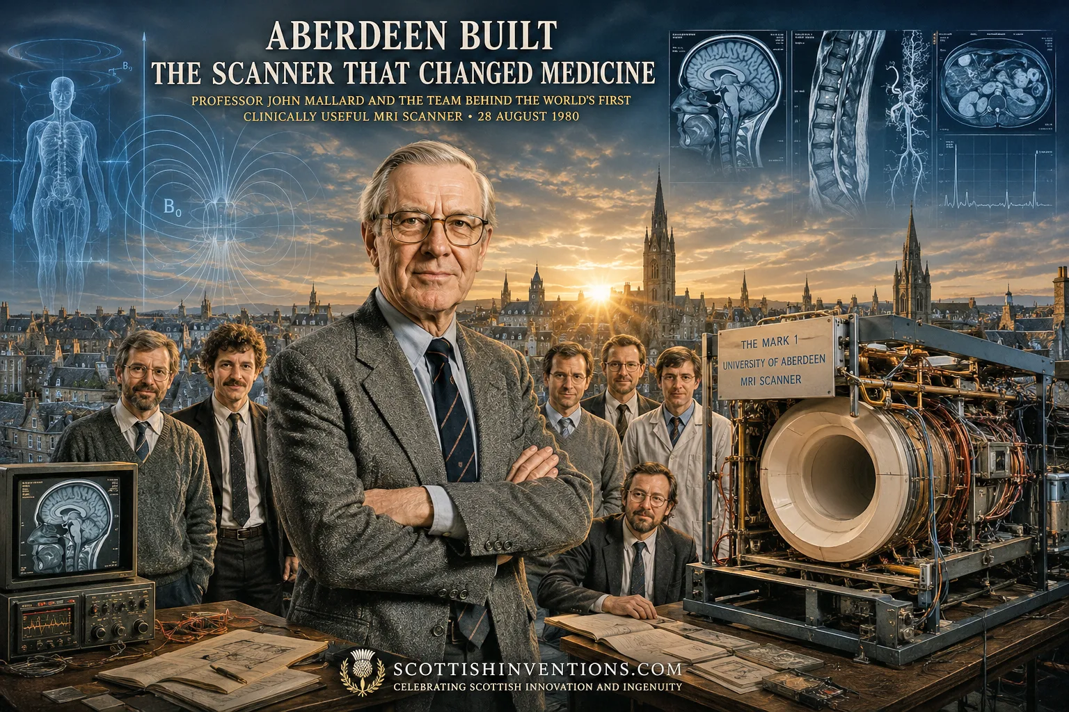
American physics discovered the phenomenon. An American physician proposed the medical use and produced the first human scan. An American chemist and a British physicist won the Nobel Prize for the imaging method. And on 28 August 1980, in a draughty laboratory in Aberdeen, a team of "half-mad scientists" produced the first MRI scan good enough to actually help a doctor treat a patient — using a technique still inside almost every scanner on Earth.
John Mallard · 1927–2021James Hutchison · 1938–2018University of AberdeenReading time · 22 minUpdated 2 August 2026
In Brief
No single person or country invented MRI. Felix Bloch and Edward Purcell discovered nuclear magnetic resonance in the United States in 1946. Raymond Damadian, an American physician, proposed in 1971 that NMR could detect tumours and produced the first human NMR scan in 1977. Paul Lauterbur, an American chemist, published the gradient-based imaging method in 1973; Peter Mansfield, a British physicist at Nottingham, developed the mathematics that made fast imaging practical — and the two shared the 2003 Nobel Prize in Physiology or Medicine. What a team at the University of Aberdeen, led by Professor John Mallard with James Hutchison and Bill Edelstein, contributed was spin-warp imaging, published in 1980 and still the technique inside nearly every clinical scanner today, and the world's first genuinely diagnostic whole-body MRI scan of a patient, on 28 August 1980. Aberdeen did not invent MRI outright. It invented the specific encoding method that made MRI clinically practical, and it built the first machine to prove that in a real patient.
Scottish Impact Index (SI Score)
- Global Impact
- 10/10
- Longevity
- 9/10
- Economic / Material Impact
- 9/10
- Originality / Distinctiveness
- 8/10
- Legacy (Built Upon)
- 9/10
- Recognition
- 6/10
Key Facts
- Discovery
- Spin-warp imaging (1980) — the phase-encoding technique behind almost all clinical MRI — and the first clinically useful whole-body MRI scan
- Key figures
- Professor John Mallard OBE (1927–2021); James (Jim) Hutchison (1938–2018); William (Bill) Edelstein
- Institution
- Department of Medical Physics, University of Aberdeen
- Spin-warp published
- Hutchison, Edelstein, Johnson & Redpath, Physics in Medicine and Biology, 1980
- First clinical scan
- 28 August 1980, Aberdeen Royal Infirmary — Mark 1 scanner, 0.04 T resistive magnet
- Patent
- US 4,506,222 (Hutchison, Edelstein, Johnson, Redpath, Mallard), granted 1985; held by NRDC/BTG
- Rival claim — physics
- Felix Bloch & Edward Purcell discover nuclear magnetic resonance, 1946 (Nobel Prize in Physics, 1952)
- Rival claim — medical concept
- Raymond Damadian (US), 1971 Science paper on tumour relaxation times; 1977 'Indomitable' first human NMR scan
- Rival claim — imaging method
- Paul Lauterbur (US), gradient-based image formation, Nature, 1973; Peter Mansfield (UK), echo-planar mathematics — jointly awarded the 2003 Nobel Prize in Physiology or Medicine
- Global impact today
- Roughly 50,000 MRI systems installed worldwide; on the order of 95 million scans performed annually
- Claim status
- DISPUTED — genuinely shared credit. Aberdeen did not invent MRI outright; it invented spin-warp encoding and built the first machine that produced diagnostic-quality clinical images
The Physics Behind MRI
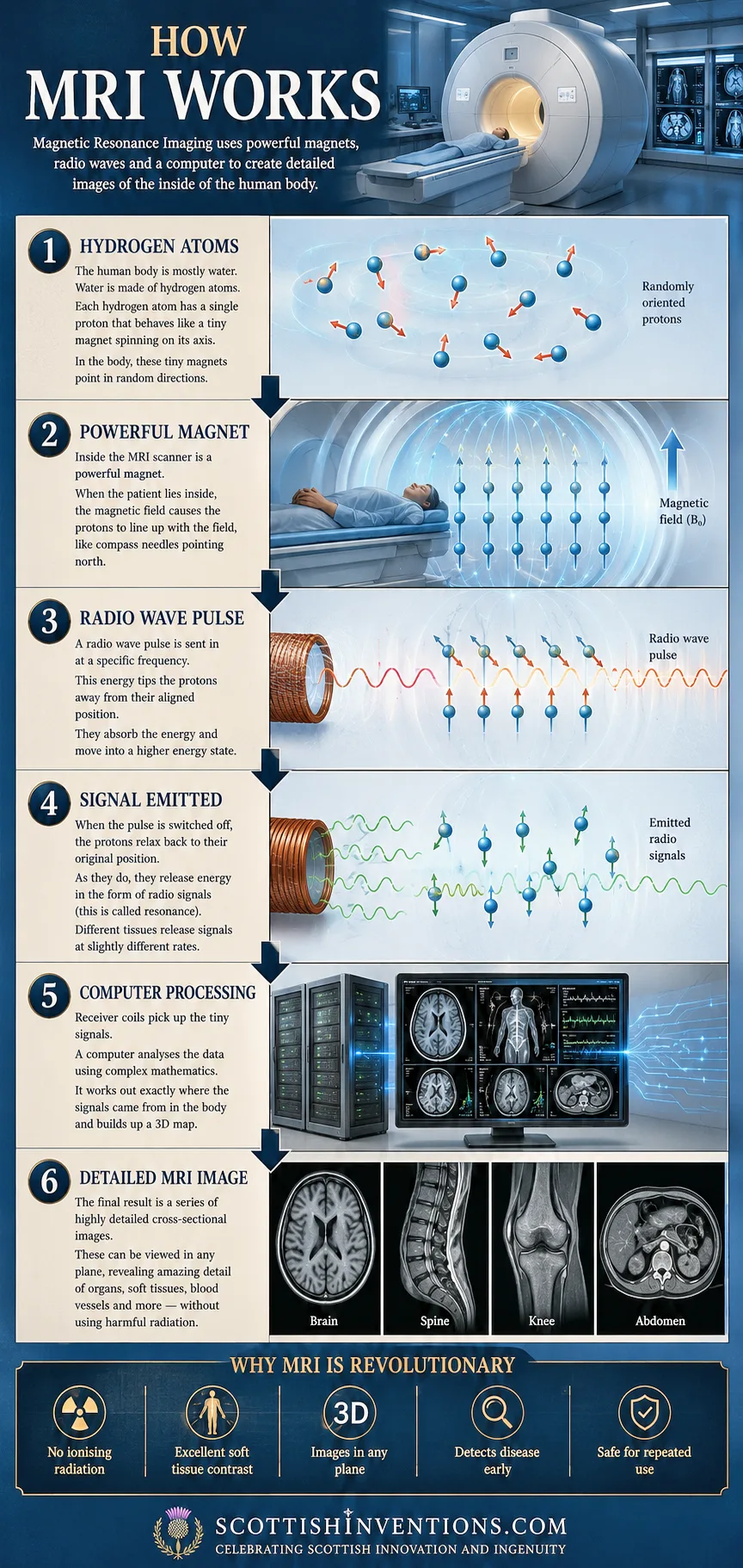
The body is mostly water, and water is full of hydrogen. The nucleus of a hydrogen atom is a single proton, and a proton behaves like a tiny spinning magnet. Scattered randomly, these countless tiny magnets point every which way. Place a person inside the powerful, uniform magnetic field of an MRI scanner, and a small fraction of them line up, like compass needles finding north.
The scanner then fires a precisely tuned pulse of radio waves, which knocks the aligned protons out of position. As the pulse switches off, the protons relax back into alignment — and as they do, each emits a faint radio signal of its own. Different tissues, containing different amounts of water bound in different ways, relax and emit at slightly different rates. A receiver coil picks up this whisper of signal, and a computer turns it into an image.
The hard problem was never detecting that signal — chemists had used nuclear magnetic resonance (NMR) as an analytical tool since the late 1940s. The hard problem was working out where in the bodyeach part of the signal had come from, so that a two-dimensional or three-dimensional picture could be reconstructed. That is the problem every contributor in this story — Lauterbur, Mansfield, Damadian and the Aberdeen team — was, in different ways, trying to solve. As Mallard told the BBC in 2018: "we had X-rays that were telling us everything about bones, but we had absolutely nothing that was telling us about the soft wet tissues within the body. And that's what MRI did."
The Road to MRI: No Single Inventor
Great inventions rarely have one parent, and MRI is a textbook case — arguably the clearest case in this entire collection. Felix Bloch at Stanford and Edward Purcell at Harvard independently discovered nuclear magnetic resonance in 1946, work that earned them the shared 1952 Nobel Prize in Physics. For the next quarter-century, NMR was a laboratory technique used by chemists to study the structure of molecules — nobody was imaging a human being with it, and nobody thought it obviously would.
Turning a chemistry tool into a medical instrument required at least three further, largely independent leaps: a reason to believe NMR could detect disease in a living body; a mathematical method for converting NMR signals into a spatial image; and an engineering effort to build a machine reliable enough to be used, repeatedly, on real patients in a hospital. Those three leaps were made, in turn, by an American physician, an American chemist working with a British physicist, and a Scottish university department. Each is treated fully below, in the order the contributions were made.
Damadian's Indomitable: The Medical Leap
Raymond Damadian, an American physician and biophysicist, made the pivotal medical connection. In a 1971 paper in Science, he reported that excised cancerous tissue exhibited significantly different NMR relaxation times (T1 and T2) from healthy tissue — the first solid evidence that NMR might detect disease non-invasively inside a living body, rather than merely describe molecular structure in a test tube. This was the idea that made everything that followed worth attempting.
Damadian went further than proposing the idea: in 1977, his team produced the first NMR scan of a living human body, using a machine he had built and named "Indomitable." The scan — of his colleague Larry Minkoff's chest — took roughly five hours and assembled an image from 106 separate points. It was slow, of low resolution by any later standard, and impractical as a clinical tool, but it was a genuine first: proof that a whole human torso could be imaged by magnetic resonance at all.
Damadian's technique used a point-by-point field-focusing method rather than the gradient-based image formation that became the industry standard, and this is one reason historians give for why the Nobel committee later credited Lauterbur and Mansfield rather than Damadian for "MRI" specifically. Damadian disagreed forcefully with that judgement, and his exclusion from the 2003 Nobel Prize remains the most publicly bitter dispute in this entire history — treated fully below.
Lauterbur and Mansfield: The Imaging Method
Paul Lauterbur, an American chemist at the State University of New York, Stony Brook, made the breakthrough that is generally regarded as the founding method of MRI image formation. In Nature on 16 March 1973, he showed that superimposing a deliberate, controlled magnetic field gradient onto the main magnetic field let researchers determine precisely where in an object an NMR signal originated — the essential principle that makes image reconstruction possible. He called his technique "zeugmatography."
Peter Mansfield, a physicist at the University of Nottingham, developed the mathematical framework for analysing the signals produced by such gradients and, crucially, invented echo-planar imaging (EPI), a method that could acquire images dramatically faster than earlier techniques — fast enough, eventually, to image a beating heart. Lauterbur and Mansfield jointly received the 2003 Nobel Prize in Physiology or Medicine "for their discoveries concerning magnetic resonance imaging."
It was against this backdrop — a workable underlying physics from the Americans in 1946, a medical rationale and first human scan from Damadian in 1971 and 1977, and a founding imaging method from Lauterbur and Mansfield from 1973 onward — that John Mallard's team in Aberdeen took on the separate, stubbornly practical problem of building a scanner that could be used, reliably and repeatedly, on real patients in a working hospital.
Mallard's Aberdeen Department
John Rowland Mallard was born in 1927 in Kingsthorpe, Northampton, and completed a PhD on the magnetic properties of uranium at University College, Nottingham. He built his early career in medical physics at Hammersmith Hospital in London, where in 1959 he constructed the first whole-body isotope scanner in Britain. In 1964 he published a paper in Nature suggesting that magnetic resonance might one day diagnose cancer — a paper that, at the time, went largely unnoticed.
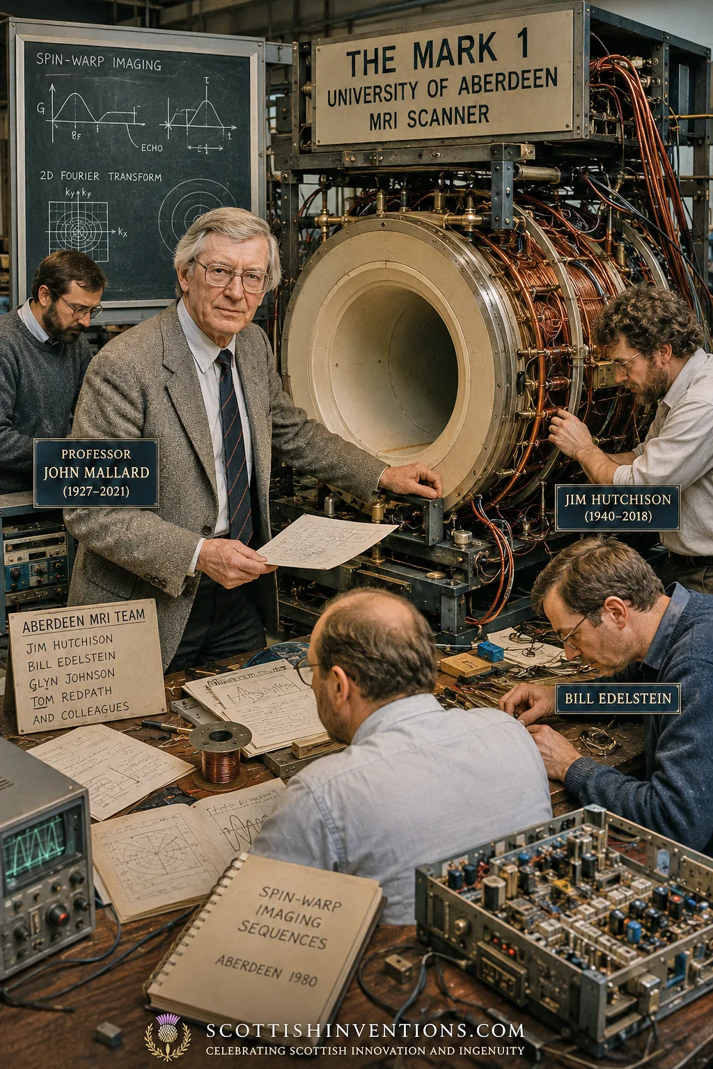
In 1965 Mallard was appointed to the newly created Chair of Medical Physics at the University of Aberdeen — the first such chair in Scotland. He appointed James (Jim) Hutchison to pursue magnetic resonance and, over the following years, built a team that came to include Hutchison, Bill Edelstein, Glyn Johnson and Tom Redpath. The work was relentlessly improvised: in 1974 Mallard and Hutchison obtained the first MRI image of a live mouse on a small desktop apparatus they had built themselves; in 1975 they imaged a dead mouse, the first pathology ever imaged by magnetic resonance.
After what Mallard described as "a struggle," he persuaded the UK's Medical Research Council to grant around £30,000 to build a whole-body scanner, the Mark 1. Its 0.04-tesla resistive magnet was built by Oxford Instruments to Hutchison's design and delivered in 1977. The team constructed the rest themselves — radiofrequency coils wound from copper pipe bought at a local hardware store, and gradient coils wound on a discarded plastic tube salvaged from a nearby park's playground.
But early images produced by line-scanning techniques — Aberdeen's own approach and, in different forms, those of most other groups worldwide — suffered from severe motion artefacts. The team's own candid word for them was "blobby." They were not of diagnostic quality, and everyone working in the field in the late 1970s knew it.
Spin-Warp Imaging, 1980
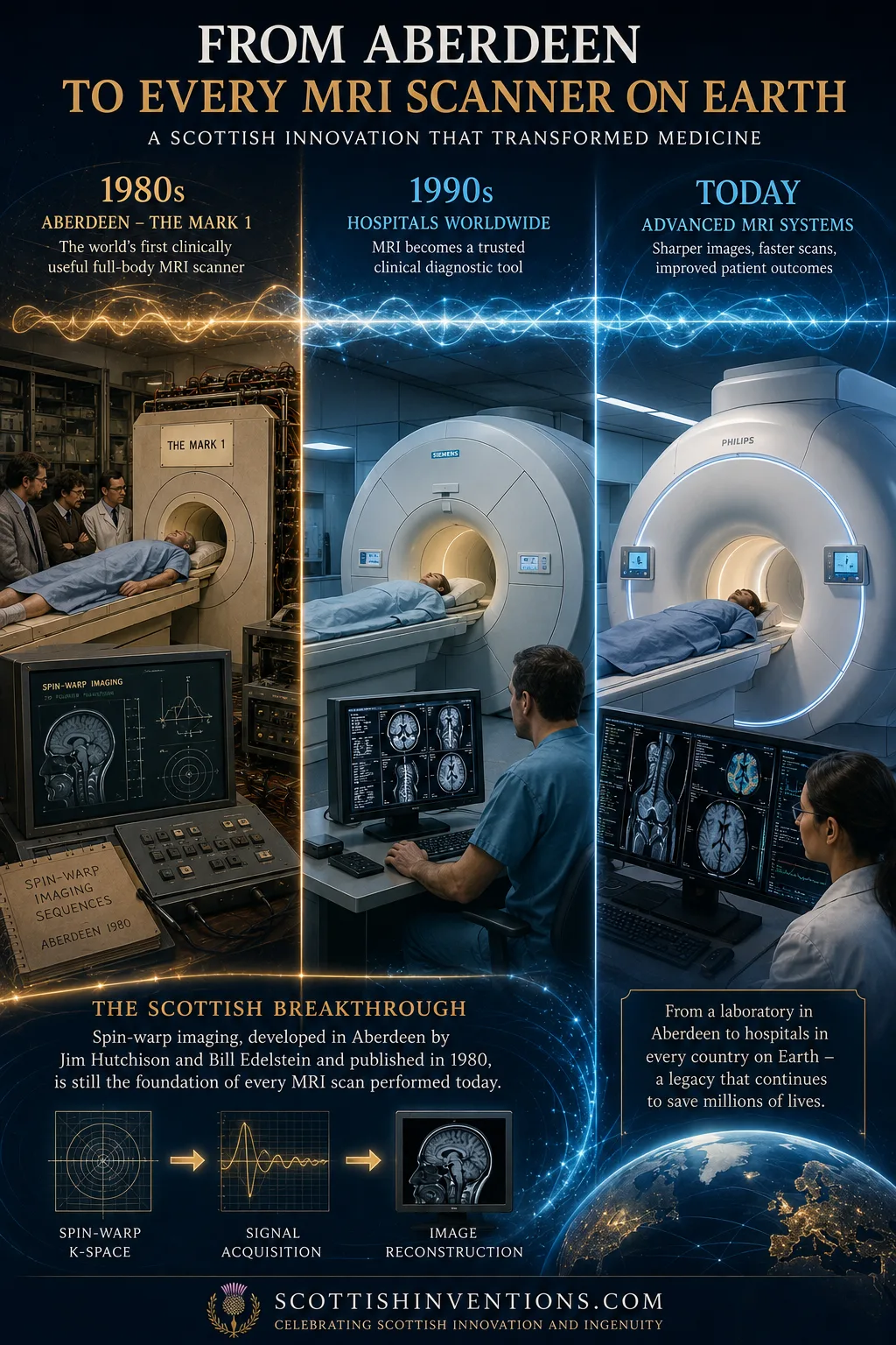
The breakthrough that solved the blobby-image problem came in 1980, when Jim Hutchison and Bill Edelstein developed spin-warp imaging, published with Glyn Johnson and Tom Redpath as "Spin warp NMR imaging and applications to human whole-body imaging" in Physics in Medicine and Biology. Spin-warp used a variable-amplitude phase-encoding magnetic gradient — applied in incremental steps rather than continuously — which proved far more tolerant of magnet imperfections and patient movement than the line-scanning and simple back-projection methods used until then, including those used by Lauterbur's and Damadian's groups.
The practical effect was transformative: spin-warp produced stable, reproducible, largely artefact-free images of genuine diagnostic quality in a matter of minutes, rather than the hours that Damadian's 1977 "Indomitable" scan had required. This is the specific, narrow, well-documented claim this article makes on Aberdeen's behalf: not that Aberdeen discovered magnetic resonance, and not that Aberdeen invented the idea of medical imaging with it, but that Aberdeen solved the encoding problem in the form that stuck. As Professor David Lurie of the University of Aberdeen has summarised it, spin-warp imaging "is still used by every single MRI scanner in the world today."
The First Patient Scan — 28 August 1980
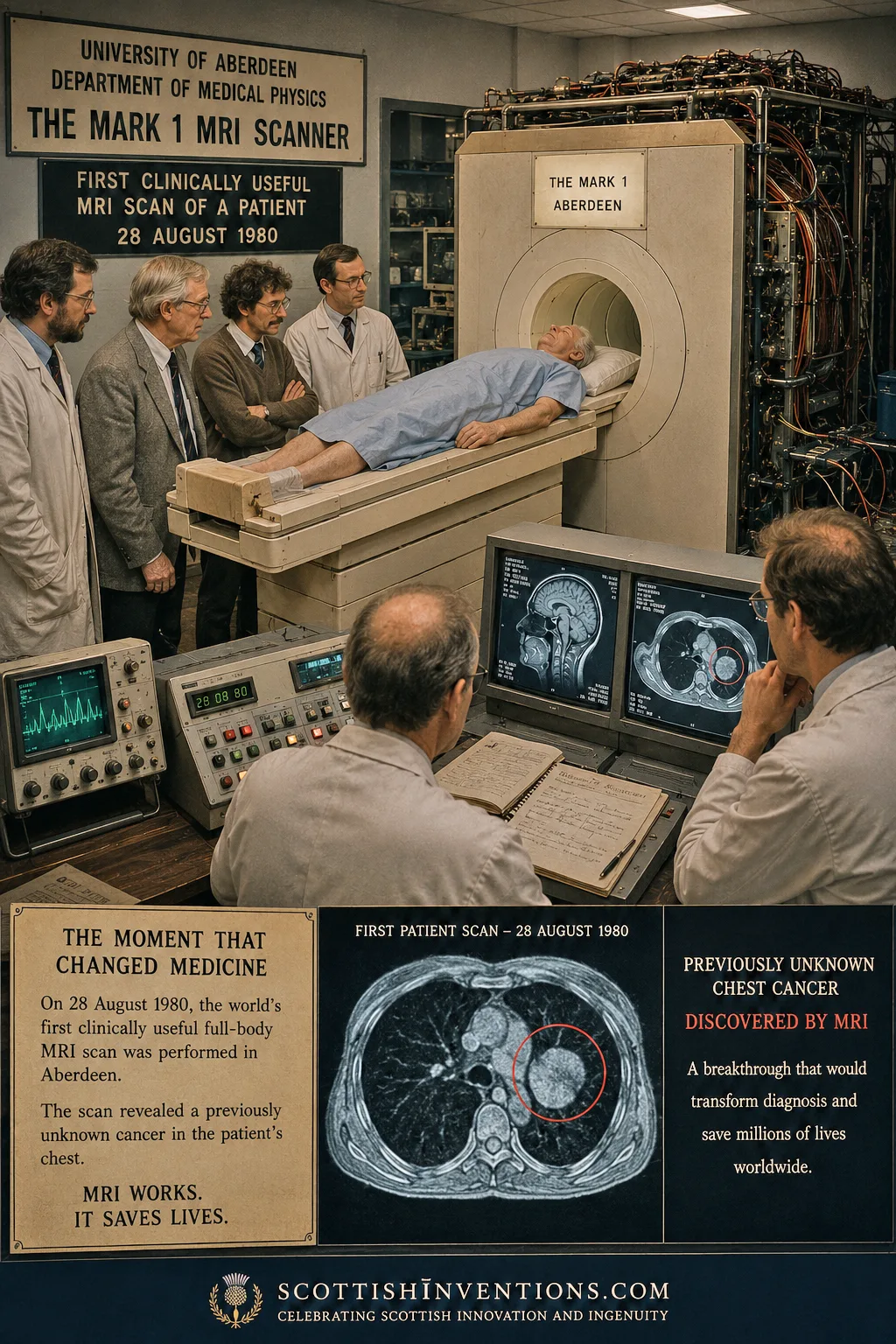
Then came the moment that justified two decades of work. On 28 August 1980, the Aberdeen team scanned their first patient: an elderly man from Fraserburgh with terminal cancer, who bravely consented to the experimental procedure. The resulting image revealed clear abnormalities in his liver — including a secondary tumour that had not previously been known about. The machine had not merely confirmed what doctors already suspected; it had found something they did not know.
The consultant radiologist who performed that scan, Dr Francis Smith, later described the development of MRI as "as important to medicine as the discovery of x-rays was in 1895." The world's first diagnostic MRI service opened at Aberdeen Royal Infirmary in 1981, and the Mark 1 went on to scan more than 1,000 patients before it was retired in 1983. It survives today, on public display at the Suttie Arts Space, Aberdeen Royal Infirmary.
In Mallard's own words
In a 2006 paper in Physics in Medicine and Biology, John Mallard reflected candidly on the limits of recognition his team had received, observing that popular accounts of MRI "tended to imply it was invented in America, when all the early developmental work was done in this country." His complaint was specifically about the engineering and clinical work — not a claim to have discovered the physics or the medical concept, both of which he consistently attributed to Bloch, Purcell and Damadian.
The spin-warp patent, US 4,506,222, was held by the UK's National Research Development Corporation, later the British Technology Group, which licensed the Aberdeen technology to most of the major manufacturers, including General Electric and Hitachi — a licensing arrangement that helped underpin the global MRI industry. Bill Edelstein himself moved to GE's Corporate Research and Development Center in Schenectady, New York, in 1980, where he helped establish an MRI group within the company, carrying the Aberdeen method directly into the world's largest medical-imaging manufacturer.
The 2003 Nobel Controversy
The 2003 Nobel Prize in Physiology or Medicine went to Paul Lauterbur and Peter Mansfield "for their discoveries concerning magnetic resonance imaging." The award was immediately controversial, and for two distinct reasons that are often run together but should be kept separate.
The louder controversy concerned Raymond Damadian, who was excluded despite his 1971 paper and 1977 "Indomitable" scan. Damadian, furious, took out full-page advertisements in the Washington Post, the New York Times and the Los Angeles Times, headlined "The Shameful Wrong That Must Be Righted," at an estimated cost of some $290,000. The Nobel committee does not explain its deliberations publicly, and the dispute has never been formally resolved; this site records both the scale of Damadian's contribution and the fact of his exclusion without adjudicating between them.
The quieter controversy concerned Aberdeen. Mallard, Hutchison and Edelstein — whose spin-warp method had by 2003 become the near-universal standard, and whose 1980 scanner had produced the first clinically useful clinical images years before most rival groups achieved comparable results — were likewise passed over. As a tribute in the Journal of Magnetic Resonance Imaging later put it, without the Aberdeen group's contribution "MR might have remained a curiosity in the engineering labs for a long while." Neither Mallard nor Hutchison mounted anything like Damadian's public campaign; both were, by most accounts, notably reticent about claiming personal credit.
Legacy: Mark 1 to the Modern World
MRI is now one of the most important diagnostic tools in medicine, used to investigate cancer, neurological disease, the spine, joints, the heart and much else besides. Industry estimates from GE HealthCare and others put the global installed base at roughly 50,000 MRI systems, performing on the order of 95 million scans a year worldwide — with the United States alone accounting for close to 40 million scans annually, and Japan reporting the highest density of scanners per head of population anywhere in the world.
Aberdeen's own Mark 1 scanner, after its retirement from front-line clinical service in 1983, continued to be of documented interest internationally as the underlying spin-warp technology was licensed by Britain's NRDC/BTG to major manufacturers and adapted into successive generations of commercial scanners; Aberdeen-derived scanner designs and Aberdeen-trained expertise subsequently found their way into clinical services well beyond the UK, including installations in parts of Asia, as the technology was progressively commercialised through the 1980s. The specific machine itself remains on public display at the Suttie Arts Space, Aberdeen Royal Infirmary — a modest resting place for an instrument that changed medicine.
John Mallard's own honours were considerable: OBE (1992), Fellow of the Royal Society of Edinburgh (1972), Fellow of the Royal Academy of Engineering (1993), and the Freedom of the City of Aberdeen (2004), an honour he shared with Nelson Mandela and Sir Alex Ferguson. He died on 25 February 2021, aged 94. Jim Hutchison, the modest and brilliant physicist at the heart of the spin-warp breakthrough, died in 2018 aged 77; colleagues recalled that he "never wanted to take personal credit for anything" and simply "wanted to produce good science." He produced some of the most consequential applied science of the twentieth century — even if the world's most famous prize never quite said so.
Myth vs Evidence
Myth: "MRI was invented in Scotland."
Evidence: The underlying physics (Bloch and Purcell, USA, 1946), the medical concept and first human scan (Damadian, USA, 1971 and 1977) and the Nobel-recognised imaging method (Lauterbur, USA, 1973; Mansfield, England, from the 1970s) all predate or lie outside Aberdeen's contribution. What Scotland can properly claim is spin-warp imaging (1980) and the first clinically useful whole-body scan.
Myth: "Damadian was cheated of the Nobel Prize, full stop."
Evidence: This is genuinely disputed, not settled. Damadian's 1971 insight and 1977 scan were real firsts, but his point-by-point scanning method differed from the gradient-imaging approach the Nobel committee specifically credited to Lauterbur and Mansfield. The committee does not publish its reasoning; historians remain divided.
Myth: "Aberdeen's scanner was just an engineering copy of others' ideas."
Evidence: Spin-warp imaging was a genuine, published, patented technical innovation — not a copy of Lauterbur's or Damadian's methods — and it is the specific encoding approach the industry adopted, worldwide, as its standard.
Did You Know?
- The Aberdeen team built early gradient coils by winding wire onto a discarded plastic tube salvaged from a children's playground.
- Damadian's 1977 human NMR scan, on a machine called 'Indomitable', took roughly five hours to produce a single low-resolution image.
- Spin-warp imaging, published in Aberdeen in 1980, is still the technique inside almost every MRI scanner made today.
- The first Aberdeen patient scan, on 28 August 1980, found a secondary cancer doctors did not already know about.
- Raymond Damadian spent an estimated $290,000 on newspaper advertisements protesting his 2003 Nobel exclusion.
- John Mallard was given the Freedom of the City of Aberdeen in 2004, an honour he shared with Nelson Mandela and Sir Alex Ferguson.
Timeline
1946
Felix Bloch and Edward Purcell independently discover nuclear magnetic resonance
The physical phenomenon underlying MRI; they share the 1952 Nobel Prize in Physics
1959
John Mallard builds the UK's first whole-body isotope scanner at Hammersmith Hospital, London
Establishes Mallard's career in medical imaging before he ever touches magnetic resonance
1964
Mallard publishes in Nature suggesting NMR might diagnose cancer
The paper attracts almost no attention at the time
1965
Mallard appointed to the first Chair of Medical Physics in Scotland, University of Aberdeen
He begins assembling the team, including Jim Hutchison
1971
Raymond Damadian publishes in Science that tumours give different NMR relaxation signals from healthy tissue
The paper that first proposed NMR as a cancer-detection tool in living tissue
1973
Paul Lauterbur publishes in Nature the use of magnetic field gradients to localise NMR signals and form images
The foundational method of image formation; later shared the 2003 Nobel Prize
1974
Mallard and Hutchison obtain the first MRI image of a live mouse in Aberdeen
On a small desktop apparatus, using line-scanning methods
1977
Damadian's team produce the first NMR scan of a living human, the five-hour 'Indomitable' chest scan
A landmark human proof of concept, though of very low resolution and impractically slow
1977
Peter Mansfield's Nottingham group develops mathematics for faster image reconstruction
Contributes to what becomes echo-planar imaging
1980
Hutchison and Edelstein publish spin-warp imaging; on 28 August the Aberdeen Mark 1 scans its first patient
The technique still used by nearly all clinical scanners; the scan reveals an unsuspected secondary cancer
1981
The world's first diagnostic MRI service opens at Aberdeen Royal Infirmary
Routine clinical use begins
1983
The Mark 1 is retired after scanning over 1,000 patients
Spin-warp is licensed by Britain's NRDC/BTG to GE, Hitachi and others
2003
The Nobel Prize in Physiology or Medicine goes to Paul Lauterbur and Peter Mansfield
Damadian and the Aberdeen team are not included; both react publicly
Today
Around 95 million MRI scans performed worldwide each year
The overwhelming majority use spin-warp phase-encoding
Notes on the Evidence
The Nobel committee gives no public reasoning. The Nobel Foundation does not disclose its full deliberations for fifty years, so all discussion of why Damadian and Aberdeen were excluded from the 2003 prize is necessarily inference from public statements, subsequent commentary and the official citation's specific wording ("discoveries concerning magnetic resonance imaging"), not from an official account of the committee's reasoning.
Patient consent and identity. The identity of Aberdeen's first scanned patient, the Fraserburgh man of 28 August 1980, is preserved in most secondary accounts without his name, consistent with medical confidentiality; this article follows that convention.
Global scan and installation figures. Figures such as "50,000 systems" and "95 million scans a year" are industry estimates (principally from GE HealthCare publications) rather than a single authoritative census, and vary somewhat by source and year.
"Half-mad scientists." This self-deprecating phrase is drawn from retrospective accounts by and about the Aberdeen team describing the improvised, low-budget nature of the Mark 1 build, rather than from a single dated quotation.
International use of Aberdeen-derived scanners. Later commercial scanners using spin-warp encoding, including installations that saw extended use in parts of Asia, descend from the licensed technology rather than being literal copies of the Aberdeen Mark 1 hardware.
Frequently Asked Questions
Who invented the MRI scanner?
There is no single inventor, and any answer that names only one person or one country is incomplete. The underlying physics — nuclear magnetic resonance — was discovered by the Americans Felix Bloch and Edward Purcell in 1946 (Nobel Prize, 1952). The idea of using NMR to detect disease in living tissue was proposed by the American physician Raymond Damadian in 1971, who produced the first human NMR scan in 1977. The method of turning NMR signals into images using field gradients was published by the American chemist Paul Lauterbur in 1973, with further mathematical development by the British physicist Peter Mansfield; they shared the 2003 Nobel Prize. What the University of Aberdeen team, led by John Mallard with James Hutchison and Bill Edelstein, contributed was spin-warp imaging (1980) — the specific encoding technique used in nearly every clinical scanner today — and the first machine to produce genuinely useful, diagnostic-quality whole-body clinical images, on 28 August 1980.
Did Aberdeen invent MRI?
Not outright, and this site does not claim that. Aberdeen did not discover nuclear magnetic resonance (Bloch and Purcell, 1946), did not propose using it to detect tumours (Damadian, 1971), and did not publish the founding method of gradient-based image formation (Lauterbur, 1973). What Aberdeen did originate, and did first, is spin-warp imaging — the phase-encoding technique published by Hutchison and Edelstein in 1980 that is still used by virtually every MRI scanner in the world — and the first whole-body scan of a patient, on 28 August 1980, that was of genuine diagnostic quality and found real, previously unknown disease.
What exactly is spin-warp imaging, and why does it matter so much?
Spin-warp is a variant of two-dimensional Fourier-transform imaging that uses a variable-amplitude 'phase-encoding' magnetic gradient to record where each part of a signal originated in the body. Published by Jim Hutchison, Bill Edelstein, Glyn Johnson and Tom Redpath in Physics in Medicine and Biology in 1980, it is markedly more tolerant of imperfections in the magnetic field and of patient movement than the line-scanning and back-projection methods used by earlier groups, including Lauterbur's and Damadian's. That robustness is precisely why manufacturers adopted it: as Professor David Lurie of the University of Aberdeen has put it, spin-warp imaging 'is still used by every single MRI scanner in the world today.'
What did Raymond Damadian actually do, and was he treated unfairly?
Damadian's 1971 paper in Science was the first to show that excised cancerous tissue had measurably different NMR relaxation times from healthy tissue — the medical insight that made NMR imaging worth pursuing at all. In 1977 his team produced the first NMR scan of a living human body, a gruelling five-hour, 106-point scan of his colleague Larry Minkoff's chest, on a machine nicknamed 'Indomitable.' Damadian was excluded from the 2003 Nobel Prize, which recognised Lauterbur and Mansfield for the image-formation methods rather than Damadian's earlier diagnostic concept and scanning method, which used a point-by-point technique rather than the gradient-based imaging that became standard. Damadian responded with full-page newspaper advertisements headlined 'The Shameful Wrong That Must Be Righted.' The Nobel committee does not explain its reasoning publicly, and reasonable historians of science continue to disagree about the fairness of the omission; both perspectives are recorded here without this site taking sides in the dispute.
Why did Lauterbur and Mansfield win the 2003 Nobel Prize instead of Damadian or the Aberdeen team?
The Nobel Prize in Physiology or Medicine specifically recognised 'discoveries concerning magnetic resonance imaging' — that is, the general scientific principles for converting NMR signals into images, credited to Lauterbur's 1973 gradient method and Mansfield's mathematical and technical refinements, including echo-planar imaging. The Nobel committee's citation did not extend to either Damadian's earlier diagnostic proposal and scanning approach or to Aberdeen's later, and arguably more clinically decisive, engineering achievement of spin-warp imaging and the first genuinely useful whole-body scanner. The Nobel Foundation does not comment on why particular contributors were excluded, so the omissions of both Damadian and Aberdeen remain matters of informed dispute rather than settled fact.
What happened on 28 August 1980 in Aberdeen?
The Aberdeen Mark 1 scanner, built on a shoestring budget by Mallard's team, produced its first clinical scan of a patient — an elderly man from Fraserburgh with terminal cancer, who consented to the experimental procedure. The image showed clear abnormalities in his liver, including a secondary tumour that had not previously been detected. It was the first time a whole-body MRI scan had produced clinically useful diagnostic information about a real patient, rather than a slow, low-resolution research image.
What is the Mark 1 scanner, and what happened to it?
The Mark 1 was Aberdeen's first whole-body scanner, built around a 0.04-tesla resistive magnet made by Oxford Instruments to Jim Hutchison's design, with radiofrequency coils improvised from copper pipe and gradient coils wound on a discarded plastic tube. It scanned more than 1,000 patients before being retired in 1983, when the world's first diagnostic MRI service, opened at Aberdeen Royal Infirmary in 1981, moved to newer equipment. The Mark 1 itself survives on public display at the Suttie Arts Space, Aberdeen Royal Infirmary. Later Aberdeen-designed and Aberdeen-derived scanners went on to see extended clinical service abroad, including installations in parts of Asia, as the technology was progressively commercialised and licensed worldwide.
How many MRI scans are performed today, and how central is the Aberdeen method?
Industry estimates (GE HealthCare and others) put the global installed base at roughly 50,000 MRI systems, performing on the order of 95 million scans a year. The overwhelming majority of these machines, regardless of manufacturer, use some form of phase-encoded Fourier-transform imaging descended directly from the spin-warp method Hutchison and Edelstein published in 1980.
Is it fair to call this a 'Scottish invention' given how many other countries were involved?
Only with precision. MRI as a whole is an international achievement built on American physics (Bloch, Purcell), an American medical insight and early human scan (Damadian), an American gradient-imaging method (Lauterbur) and British mathematical and technical work at Nottingham (Mansfield) as well as Aberdeen. What can fairly be called a specifically Scottish contribution is spin-warp imaging, developed and published at the University of Aberdeen in 1980, and the world's first clinically useful whole-body diagnostic scan, performed there on 28 August 1980. This page presents that narrower, defensible claim rather than an unqualified assertion that MRI was 'invented in Scotland.'
Sources & Further Reading
Tier 1 · Primary
- Hutchison, J. M. S., Edelstein, W. A., Johnson, G., Redpath, T. W. & Mallard, J. R. — US Patent 4,506,222, "Imaging Systems," granted 1985.
Tier 2 · Scholarly and institutional
- Bloch, F. — "Nuclear Induction," Physical Review, 70 (1946); Purcell, E. M., Torrey, H. C. & Pound, R. V. — "Resonance Absorption by Nuclear Magnetic Moments in a Solid," Physical Review, 69 (1946).
- Damadian, R. — "Tumor Detection by Nuclear Magnetic Resonance," Science, 171 (1971), pp. 1151–1153.
- Lauterbur, P. C. — "Image Formation by Induced Local Interactions: Examples Employing Nuclear Magnetic Resonance," Nature, 242 (1973), pp. 190–191.
- Mansfield, P. & Grannell, P. K. — "NMR 'Diffraction' in Solids?," Journal of Physics C, 6 (1973).
- Hutchison, J. M. S., Edelstein, W. A., Johnson, G. & Redpath, T. W. — "Spin Warp NMR Imaging and Applications to Human Whole-Body Imaging," Physics in Medicine and Biology, 25 (1980), pp. 751–756.
- Mallard, J. R. — "Fond Memories of Physics in Medicine and Biology During the First 20 Years," Physics in Medicine and Biology, 51 (2006).
- The Nobel Foundation — "The Nobel Prize in Physiology or Medicine 2003," official citation and press release, Nobelprize.org.
- Filler, A. G. — "The History, Development and Impact of Computed Imaging in Neurological Diagnosis and Neurosurgery: CT, MRI and DTI," Nature Precedings, 2009 (covers the Damadian/Nobel dispute in detail).
- University of Aberdeen — Department of Biomedical Physical Sciences, institutional history of the Mark 1 scanner and the spin-warp discovery.
- GE HealthCare — published estimates of global MRI installed base and annual scan volumes.
Tier 3 · General reference
- BBC News and BBC Scotland — obituaries and retrospective coverage of John Mallard and James Hutchison, 2018 and 2021.