MRI Scanner
by John Mallard & James Hutchison
Aberdeen Owns the MRI Story
When you next slide into the tunnel of an MRI scanner, spare a thought for a draughty laboratory in Aberdeen and a self-described 'small team of half-mad scientists.' The physics that makes MRI possible was discovered in the United States. The Nobel Prize for MRI went to an American and an Englishman. But the machine that first proved magnetic resonance could see inside a living person and find disease — the machine that began the modern era of medical imaging — was built in the north-east of Scotland.
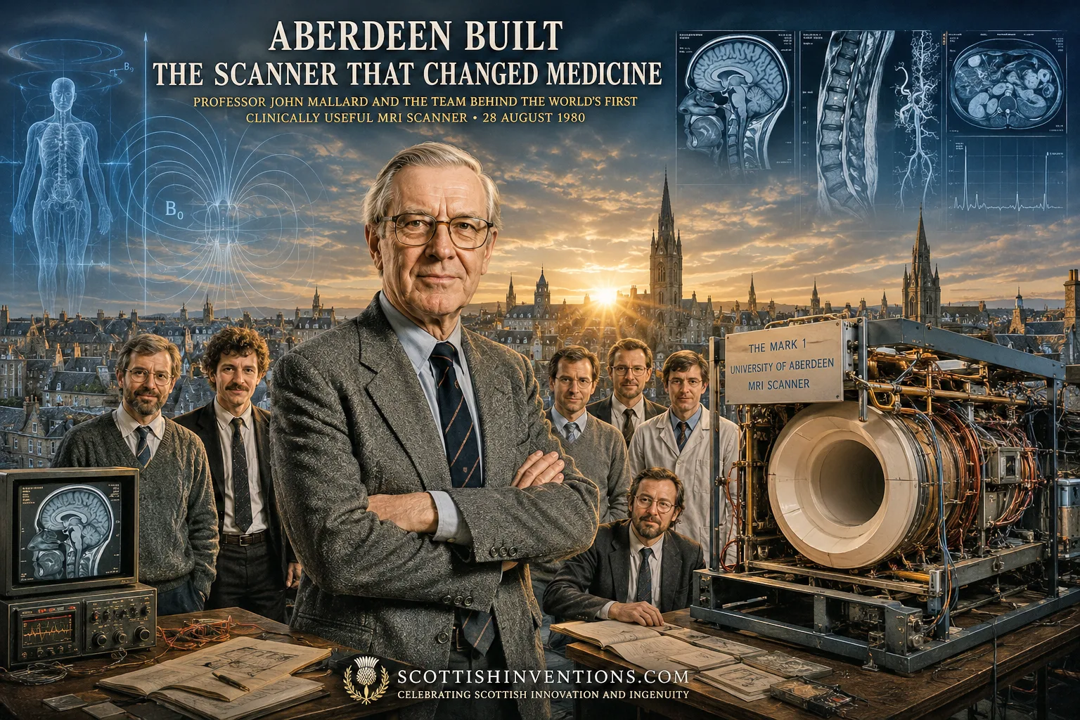
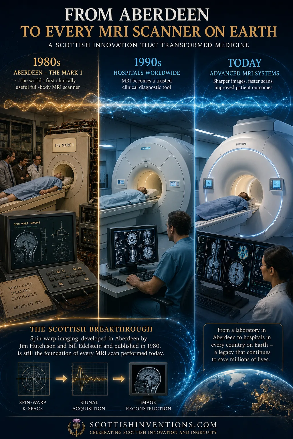
The claim that 'Aberdeen owns the MRI story' is not Scottish bravado. It is a precise, defensible historical statement, and it rests on two pillars: Professor John Mallard's team built the world's first full-body MRI scanner capable of clinically useful diagnostic images, and Jim Hutchison and Bill Edelstein invented the spin-warp imaging technique that remains the foundation of virtually all modern MRI scans.
The underlying physics of Nuclear Magnetic Resonance was worked out by Americans Felix Bloch and Edward Purcell (Nobel Prize, 1952). The methods for turning NMR signals into images earned Paul Lauterbur and Peter Mansfield the 2003 Nobel Prize. But it was the Aberdeen team that built the first machine that actually worked as a clinical diagnostic tool, and whose spin-warp method remains the industry standard. Other teams were producing blurry images of fingers and wrists that took hours; Aberdeen produced diagnostic-quality images of the whole body in around two minutes.
The Science Behind MRI
So how does it work? The trick is that your body is mostly water, and water is full of hydrogen atoms. The nucleus of a hydrogen atom is a single proton, and a proton behaves like a tiny spinning magnet. Normally these countless tiny magnets point in random directions. But when you lie inside an MRI scanner's powerful magnet, a small fraction of them line up with the field, like compass needles.
The scanner then sends in a pulse of radio waves tuned to exactly the right frequency. This knocks the protons out of alignment. When the radio pulse switches off, the protons relax back into line — and as they do, they emit a faint radio signal of their own. That signal is the key. Different tissues — fat, muscle, healthy organs, tumours — contain different amounts of water and release their signal at slightly different rates. A receiver coil picks up these whispers, and a computer assembles them into an astonishingly detailed picture.
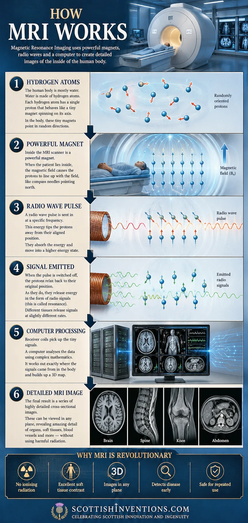
Why was this revolutionary? A conventional X-ray is superb at showing bone but poor at distinguishing the 'soft, wet tissues' of the body. A CT scan produces beautiful cross-sections, but does so using ionising radiation. MRI sees soft tissue with unrivalled clarity, uses no ionising radiation whatsoever, and can image the body in any plane in three dimensions. As Mallard told the BBC in 2018: 'we had X-rays that were telling us everything about bones, but we had absolutely nothing that was telling us about the soft wet tissues within the body. And that's what MRI did.'
How MRI Works — The Aberdeen Principle
Hydrogen protons
The body is mostly water, and each hydrogen nucleus is a single proton that behaves like a tiny spinning magnet.
Powerful magnet
Inside the scanner, a strong static magnetic field aligns a fraction of those protons like compass needles.
Radio-wave pulse
A precisely tuned radio pulse tips the protons out of alignment, lifting them into a higher energy state.
Emitted signal
As the protons relax back, they emit faint radio signals — different tissues release them at slightly different rates.
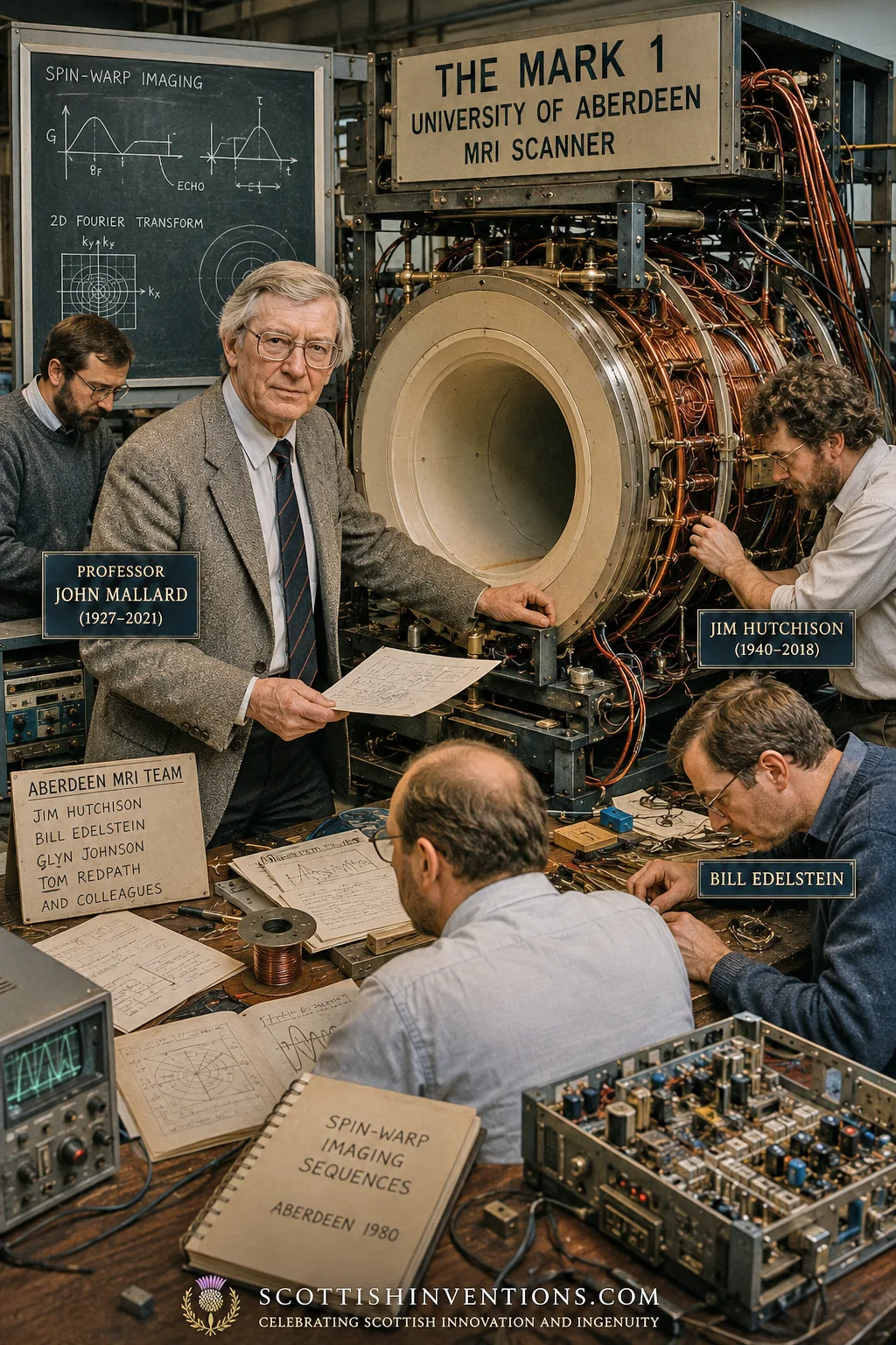
Spin-warp encoding
Hutchison and Edelstein's variable phase-encoding gradient maps each signal to its exact location, tolerant of motion and field imperfections.
Image reconstruction
A computer applies a 2D Fourier transform to assemble the signals into a high-resolution, multi-plane image — without any ionising radiation.
The Road to MRI: The Key Predecessors
Great inventions rarely have a single parent, and MRI is a textbook case. Felix Bloch and Edward Purcell independently discovered nuclear magnetic resonance in 1946, sharing the 1952 Nobel Prize in Physics. For two decades, NMR was a tool for chemists studying molecules, not doctors examining patients.
Raymond Damadian, an American physician, made the pivotal medical leap. In a 1971 paper in Science, he reported that cancerous tissue gave different NMR signals than healthy tissue. In 1977, using a machine he christened 'Indomitable,' his team produced the first NMR scan of a living human body — a five-hour, 106-point image of his assistant Larry Minkoff's chest.
Paul Lauterbur, an American chemist, made the breakthrough that turned NMR into a picture. In Nature on 16 March 1973 he showed that a deliberate magnetic field gradient let you work out where a signal came from — the essential principle of MRI image formation. Peter Mansfield, a physicist at the University of Nottingham, developed the mathematics for rapidly converting signals into images. Lauterbur and Mansfield shared the 2003 Nobel Prize.
And then there was John Mallard's team at Aberdeen, who took all of this and built the first clinically useful full-body scanner — developing spin-warp imaging along the way.
John Mallard and the Aberdeen Team in Detail
John Rowland Mallard was born in 1927 in Kingsthorpe, Northampton, and completed a PhD on the magnetic properties of uranium at University College, Nottingham. He built his early career in medical physics at Hammersmith Hospital in London, where in 1959 he built the first whole-body isotope scanner in the UK. In 1964 he published a paper in Nature suggesting magnetic resonance might be used to diagnose cancer — a paper that went largely unnoticed.
In 1965 Mallard was appointed to the chair of Medical Physics at the University of Aberdeen — the first such chair in Scotland. He arrived with a vision. He appointed Jim Hutchison to pursue magnetic resonance, and over the following years assembled a team that included Hutchison, Bill Edelstein, Glyn Johnson, Tom Redpath, and others.
The work was relentlessly improvised. In 1974 Mallard and Hutchison obtained the first MRI image of a mouse on a small desktop machine they had built; in 1975 they imaged a dead mouse, the first time pathology had been imaged by MRI. After 'a struggle,' Mallard persuaded the UK's Medical Research Council to grant £30,000 to build the Mark 1 whole-body scanner. Its 0.04-tesla resistive magnet was built by Oxford Instruments to Hutchison's design and delivered in 1977. The team built the rest themselves — using radiofrequency coils made from copper pipe bought at a hardware store and gradient coils wound on a discarded plastic tube from a local park's playground.
But there was a problem. The early images, produced by line-scanning techniques, suffered from severe motion artefacts — they were, in the team's words, 'blobby' and not of diagnostic quality. The same was true of rival groups around the world. The breakthrough came in 1980, with spin-warp imaging, developed by Hutchison and Edelstein and published as 'Spin warp NMR imaging and applications to human whole-body imaging' in Physics in Medicine and Biology. Spin-warp used a variable-amplitude phase-encoding magnetic gradient and was far more tolerant of imperfections than earlier methods, producing stable, reproducible, artefact-free images of genuine diagnostic quality in around two minutes rather than hours.
The First Patient Scan — 28 August 1980
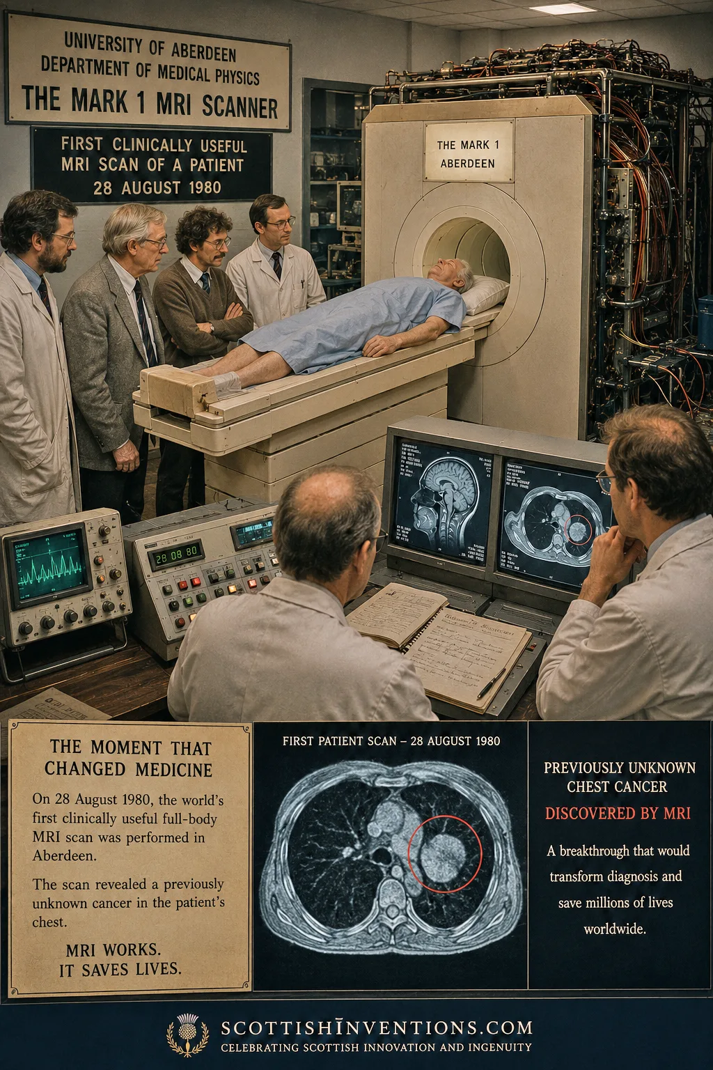
Then came the moment that justified two decades of work. On 28 August 1980, the team scanned their first patient — an elderly man from Fraserburgh with terminal cancer who bravely consented to the experiment. The image revealed clear abnormalities in his liver and a secondary tumour that had not previously been known. The machine had not merely confirmed what doctors knew; it had revealed something they did not.
The consultant radiologist who performed that first scan, Dr Francis Smith, later described the development of MRI as 'as important to medicine as the discovery of x-rays was in 1895.' The Mark 1 went on to scan more than 1,000 patients before being replaced in 1983, and the world's first diagnostic MRI service opened at Aberdeen Royal Infirmary in 1981. The scanner itself survives today, on display at the Suttie Arts Space in Aberdeen Royal Infirmary.
Mallard understood the significance keenly. In a 2006 paper in Physics in Medicine and Biology he reflected on the limits of recognition, observing that accounts of MRI 'tended to imply it was invented in America, when all the early developmental work was done in this country.' The spin-warp patent (US 4,506,222) was held by the UK's National Research Development Corporation, later the British Technology Group, which licensed the Aberdeen MRI technology to most of the major manufacturers — including Hitachi and General Electric — helping to underpin the global MRI industry. Bill Edelstein himself moved to GE's Corporate R&D Center in Schenectady in 1980, where he 'helped to establish an MRI group within the company.'
“Spin-warp imaging is still used by every single MRI scanner in the world today.”
Legacy and Impact
MRI is now one of the most important diagnostic tools in all of medicine, used to investigate cancer, neurological disease, the spine, joints, the heart and much more. According to GE HealthCare, around 50,000 MRI systems are installed globally and more than 95 million MRI scans are performed each year worldwide — with the United States alone accounting for nearly 40 million scans annually, and Japan leading the world in density at 57.39 MRI units per million population.
The 2003 Nobel Prize controversy remains the great asterisk in the MRI story. The prize went to Lauterbur and Mansfield. Raymond Damadian, furious at his exclusion, took out full-page advertisements in the Washington Post, New York Times and Los Angeles Times headed 'The Shameful Wrong That Must Be Righted' — at an estimated cost of $290,000. Less loudly, Mallard and the Aberdeen team were also overlooked, despite having built the first clinically useful scanner and invented the encoding method still in universal use. As a tribute in the Journal of Magnetic Resonance Imaging put it, without the Aberdeen group 'MR might have remained a curiosity in the engineering labs for a long while.'
The deepest measure of Aberdeen's legacy is this: the spin-warp technique its scientists pioneered is used in virtually every MRI scan done today. Mallard's own honours were considerable — appointed OBE in 1992, Fellow of the Royal Society of Edinburgh (1972), Fellow of the Royal Academy of Engineering (1993), and given the Freedom of the City of Aberdeen in 2004, an honour he shared with Nelson Mandela and Sir Alex Ferguson. He died on 25 February 2021, aged 94. Jim Hutchison, the brilliant and famously modest scientist at the heart of the spin-warp breakthrough, died in 2018 aged 77. He 'never wanted to take personal credit for anything' — he simply 'wanted to produce good science.' He produced some of the most consequential science of the twentieth century.
Frequently Asked Questions
Who invented the MRI scanner? The world's first clinically useful full-body MRI scanner was built at the University of Aberdeen by a team led by Professor John Mallard. On 28 August 1980 it produced the first diagnostic MRI of a cancer patient. The underlying physics of NMR was discovered by Bloch and Purcell (1946), and the 2003 Nobel Prize for image-formation methods went to Paul Lauterbur and Peter Mansfield — but Aberdeen built the first machine that actually worked as a clinical tool.
Was MRI invented in Scotland? The first clinically useful MRI scanner was built in Scotland, at the University of Aberdeen. The spin-warp imaging method developed there in 1980 by Jim Hutchison and Bill Edelstein remains the foundation of virtually every MRI scan performed today.
What is spin-warp imaging? Spin-warp is a variant of the two-dimensional Fourier transform method that uses a variable-amplitude 'phase-encoding' magnetic gradient to map signals to their location in the body. Developed in Aberdeen by Hutchison and Edelstein and published in 1980, it is far more tolerant of magnet imperfections and patient movement than earlier methods, producing stable, artefact-free diagnostic-quality images in minutes rather than hours.
Who was John Mallard? Professor John Rowland Mallard OBE (1927–2021) was Professor of Medical Physics at the University of Aberdeen from 1965 and led the team that built the world's first clinically useful full-body MRI scanner. He was appointed OBE in 1992, given the Freedom of the City of Aberdeen in 2004, and is widely regarded as one of the founding figures of clinical MRI.
What did the first MRI scan find? On 28 August 1980 the Aberdeen Mark 1 scanned an elderly patient from Fraserburgh with terminal cancer and revealed a secondary tumour that doctors had not previously detected — proving for the first time that MRI could find disease the existing imaging tools could not.
Why didn't Aberdeen win the Nobel Prize for MRI? The 2003 Nobel Prize in Physiology or Medicine went to Lauterbur and Mansfield 'for their discoveries concerning magnetic resonance imaging,' and was capped at three recipients. Many in the field believe the Aberdeen team's contribution — the first clinically useful scanner and the spin-warp encoding still used in every MRI today — was under-recognised. The Nobel Committee's deliberations remain sealed until 2053.
How many MRI scans are performed today? Approximately 95 million MRI scans are performed worldwide each year, on roughly 50,000 installed MRI systems (GE HealthCare estimate). Virtually every one of those scans uses the spin-warp encoding method invented in Aberdeen in 1980.
Where is the Mark 1 scanner today? The original Aberdeen Mark 1 scanner is preserved and on public display at the Suttie Arts Space in Aberdeen Royal Infirmary.
Related Inventions
Penicillin
Alexander Fleming · 1928
The world's first true antibiotic.
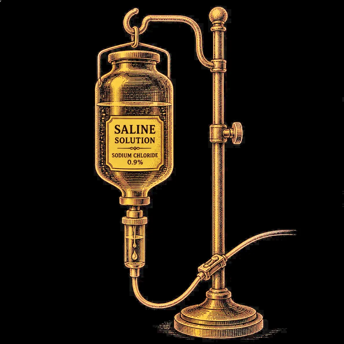
The IV Drip (Intravenous Saline)
Thomas Latta · 1832
In the spring of 1832 in Leith, Dr Thomas Latta performed the world's first intravenous saline infusion in a human being — reviving a dying cholera patient and inventing the IV drip that today saves countless lives every year.
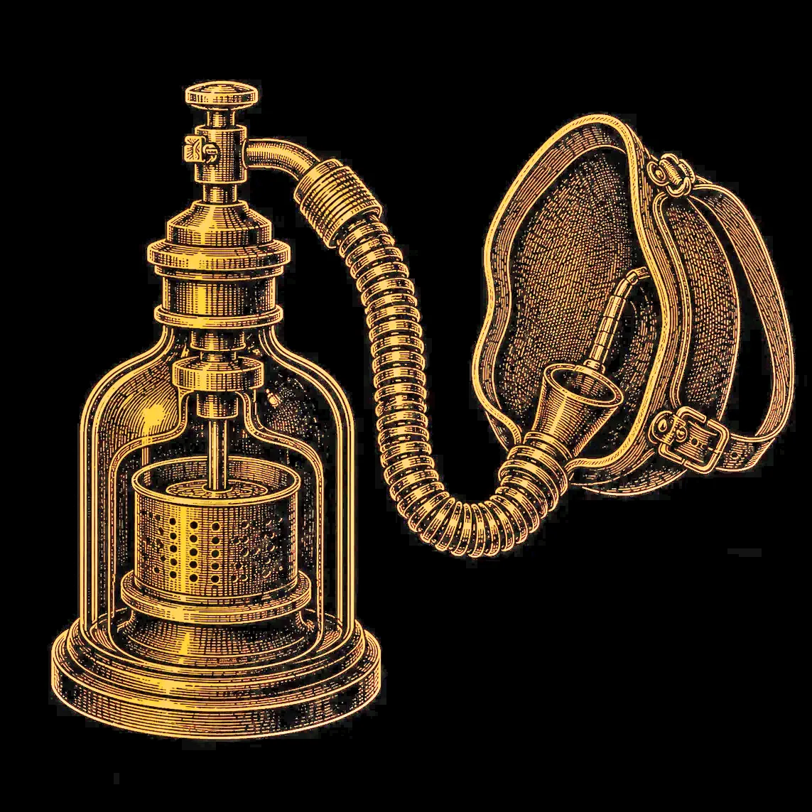
Anaesthesia / Chloroform
James Young Simpson · 1847
Surgical anaesthesia using chloroform.
Weekly Scottish Innovation Facts
Delivered to your inbox every Sunday. No spam, just brilliance.