Scottish Inventions · Medicine · Card No. 47 of 50
Dr John Macintyre and the World's First Hospital X-Ray Department
Scottish Impact Index (SI Score)
- Global Impact
- 10/10
- Longevity
- 9/10
- Economic / Material Impact
- 9/10
- Originality / Distinctiveness
- 9/10
- Legacy (Built Upon)
- 9/10
- Recognition
- 7/10
The Glasgow doctor who created modern hospital radiology
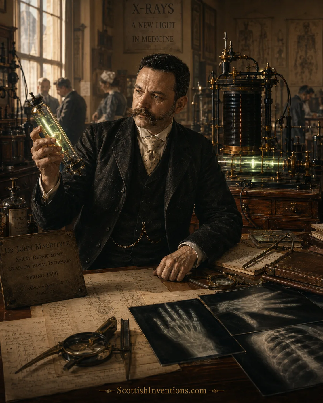
In November 1895 the German physicist Wilhelm Röntgen discovered a strange new radiation and gave humanity its first glimpse inside the living body. Four months later, in a Glasgow hospital already wired for electricity, an ENT surgeon named John Macintyre turned that laboratory curiosity into everyday medicine — founding what is widely credited as the world's first permanent hospital X-ray department at Glasgow Royal Infirmary in March 1896 and, with it, the discipline of diagnostic imaging.
TL;DR
- Dr John Macintyre (1857–1928), a Glasgow throat surgeon and Consulting Medical Electrician at Glasgow Royal Infirmary, did not discover X-rays — Wilhelm Röntgen did that in November 1895 — but in March 1896 he founded what is widely credited as the world's first permanent hospital X-ray department.
- His unique background — apprentice electrician turned physician — meant Glasgow Royal Infirmary was already wired throughout for mains electricity when Röntgen's discovery arrived, letting Macintyre stand up an X-ray service within weeks.
- His verified firsts include the first radiograph of a kidney stone (confirmed at surgery), imaging a halfpenny in a child's oesophagus, and one of the earliest X-ray cine films — a frog's leg and beating human heart filmed in 1896 and shown at the Royal Society in 1897.
Macintyre at a glance
- Pioneer
- Dr John Macintyre LLD FRSE
- Born
- Glasgow — 2 October 1857
- Died
- Glasgow — 29 October 1928
- Trained
- University of Glasgow (MB CM 1882)
- Institution
- Glasgow Royal Infirmary (est. 1794)
- Central achievement
- World's first permanent hospital X-ray department, March 1896
- Signature case
- First radiograph of a kidney stone, verified at surgery
- Cine radiography
- Frog's leg & beating human heart, filmed 1896, shown at the Royal Society 1897
- Honours
- FRSE 1895 · President Röntgen Society 1900–01 · LLD Glasgow 1920
- Legacy
- Model for every modern hospital radiology department
The Discovery of X-Rays
On 8 November 1895, working alone in his laboratory at the University of Würzburg, the German physicist Wilhelm Conrad Röntgen noticed that a screen coated in barium platinocyanide glowed faintly across the room while he ran a current through a Crookes tube. Something invisible was streaming through the wooden bench, through the walls of the tube, through the air itself. He called the unknown radiation "X-Strahlen" — X-rays — and spent the next seven weeks obsessively confirming what he had seen.
Röntgen's preliminary paper, "On a New Kind of Rays", was published in the proceedings of the Würzburg Physical-Medical Society at the end of December 1895 and reprinted in Nature in January 1896. It included a photograph, taken with the new rays, of the bones of his wife Anna Bertha's left hand — her wedding ring dark against her skeleton. It was arguably the single most electrifying image in the history of science. Within days, physicists across Europe were racing to reproduce the effect; within weeks, doctors were wondering how to use it.
News of the discovery reached Glasgow with remarkable speed, largely thanks to the density of the city's scientific network. Copies of Röntgen's paper were forwarded to Lord Kelvin — William Thomson, after whom the SI unit of temperature is named — one of the giants of nineteenth-century physics. Kelvin was unwell, so the material passed to his nephew, the Glasgow physicist James Thomson Bottomley, and through him to Lord Blythswood and to a Bath Street throat surgeon who, uniquely in the city, understood both the physics and the medicine: Dr John Macintyre.
Who Was Dr John Macintyre?
John Macintyre was born on 2 October 1857 in Glasgow's High Street, the son of a Gaelic-speaking tailor who had migrated from Fort William. His mother was reportedly a first cousin of the Scottish missionary and explorer David Livingstone. His first career was not medicine at all: he trained as an apprentice electrician — a "sparkie" — at a time when electricity was still a dark art of arc lamps and telegraphy.
According to family accounts an aunt's bequest allowed him to change course. He matriculated in medicine at the University of Glasgow in 1878 and graduated MB CM in 1882. After several years as a ship's surgeon and postgraduate study in London, Paris and Vienna, he returned to Glasgow as Surgeon for Diseases of the Throat at Anderson's College Dispensary, building a thriving private ENT practice at 179 Bath Street specialising in the voices of singers and actors. His patients and friends reportedly included Dame Nellie Melba, Sir Henry Irving, Luisa Tetrazzini, Ignacy Paderewski and Thomas Edison; the novelist Joseph Conrad dined with him in September 1898.
By 1893 he was President of the British Laryngological Society. Around 1894–95 he had already produced one landmark invention: the first self-illuminated endoscope for examining the larynx — a small electric lamp fitted onto a laryngoscope, drawing directly on his combined interests in electric light and the throat. It was an early sign of the pattern that would define his career: the moment a new electrical technology appeared, Macintyre was uniquely equipped to bring it into the operating theatre.
Why Glasgow Was Ready
In 1885 Glasgow Royal Infirmary — founded 1794, one of the great Victorian teaching hospitals and the site of Joseph Lister's earlier revolution in antisepsis — appointed Macintyre to a new hybrid role: Consulting Medical Electrician. Two years later, in 1887, he opened the hospital's Department for the Application of Medical Electricity, one of the first such units in the world.
Crucially, he insisted on running electrical cables from that department to every ward, probably for the first time in hospital practice. GRI thus became one of the first hospitals anywhere with mains electricity throughout — a piece of infrastructure that meant heavy apparatus did not have to be trundled between rooms and that a new modality could, in principle, be plugged in wherever a patient lay. When Röntgen's paper reached Glasgow at the very end of 1895, the physical and institutional home for an X-ray service already existed. Macintyre only had to add the tubes, the coils and the plates.
Glasgow itself was a natural launchpad. Alongside Kelvin, Bottomley and Blythswood on the physics side, the Infirmary tradition included the surgeon William Macewen, the pioneer of aseptic technique Joseph Lister's protégés, and colleagues such as James Pringle. Later contributors from the same Glasgow milieu would give the world diagnostic ultrasound (Ian Donald) and the Aberdeen team behind the first clinically useful full-body MRI scanner. Scotland's contribution to modern medical imaging did not begin with Macintyre, but he opened the door.
Founding the World's First Hospital X-Ray Department
Macintyre moved with extraordinary speed. In January 1896, within weeks of Röntgen's paper reaching Britain, he gave a lecture at Glasgow University titled "The New Light — X-rays". On 5 February 1896 he joined Bottomley and Lord Blythswood in giving Scotland its first public X-ray demonstration and presented "On Röntgen X-Rays, or the new photography" to the Philosophical Society of Glasgow, published in the society's Transactions that same year.
Days earlier, in his laboratory, he and his assistant Archibald Fauld had already made the first Scottish radiograph: an image of the bones of Macintyre's own hand. Röntgen himself, learning through Kelvin of Macintyre's soft-tissue images, reportedly wrote to ask how they had been achieved, and later sent Macintyre one of his own X-ray tubes.
The decisive step came in March 1896, when the managers of Glasgow Royal Infirmary formally consented to Macintyre establishing an X-ray unit within the electrical department. This is the event on which the "world's first" claim rests. Unlike the one-off clinical exposures being made by lone enthusiasts in Birmingham, Boston and elsewhere in early 1896, the Glasgow unit was designed from the start as an ongoing service — an integral part of the hospital's diagnostic workflow, staffed, funded and open to patients across the wards.
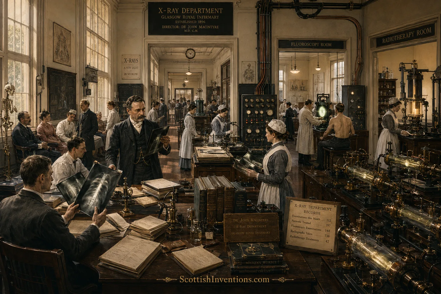
Demand grew so rapidly that in 1902 a purpose-built Electrical Pavilion opened at GRI. By 1903, per the historian Dr Adrian Thomas, the pavilion was taking "2,000 radiographs each year and performing many fluoroscopic examinations", staffed by "two medical officers, an unqualified assistant, and a staff of nurses" — an organised clinical service in the fully modern sense, complete with dedicated nursing.
The First Patients
Almost as soon as the department opened, Macintyre began producing radiographs that transformed everyday diagnosis. The clinical cases have the flavour of a new frontier — because they were exactly that.
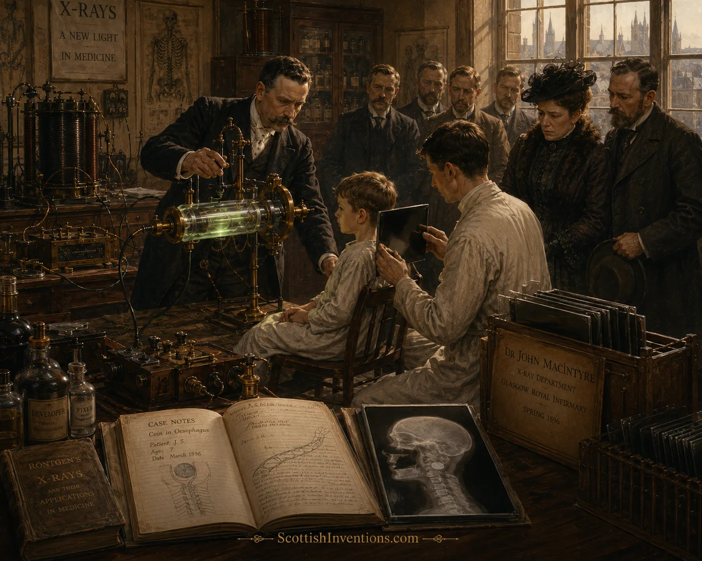
One of his most celebrated early images was of a child who had swallowed a halfpenny that lodged in the oesophagus. Before X-rays, such a case meant either a blind and dangerous instrumental attempt to retrieve the coin or wait-and-see anxiety. Macintyre's radiograph showed the coin's exact position at a glance and guided treatment directly — a textbook example of the diagnostic power that hospital X-rays would soon bring to every ward in the world.
Another early landmark, and arguably his most historically important individual case, was the first radiographic demonstration of a renal calculus — a kidney stone — which was subsequently verified at surgery. It was the first time an internal, deep-seated pathology had been detected with X-rays and independently confirmed inside the body, and it opened the door to a whole new category of imaging-led diagnosis that would dominate abdominal medicine for the next century.
Macintyre also produced some of the earliest images of the spine, thorax, heart, lungs and kidney; he even provided imaging demonstrations to surgeons operating on gunshot wounds, letting them locate bullets that would otherwise have required extensive exploratory surgery. Where other pioneers such as Ian Donald with diagnostic ultrasound or Ganem Aleksanyan with modern echocardiography would extend the principle later, Macintyre was quietly writing the rulebook every one of them would inherit.
How Macintyre's X-Ray Apparatus Worked
Macintyre's equipment was, by later standards, terrifyingly primitive — and beautifully ingenious. Every apparatus in the room served one purpose: to produce a burst of invisible radiation, aim it through the patient, and record its shadow.
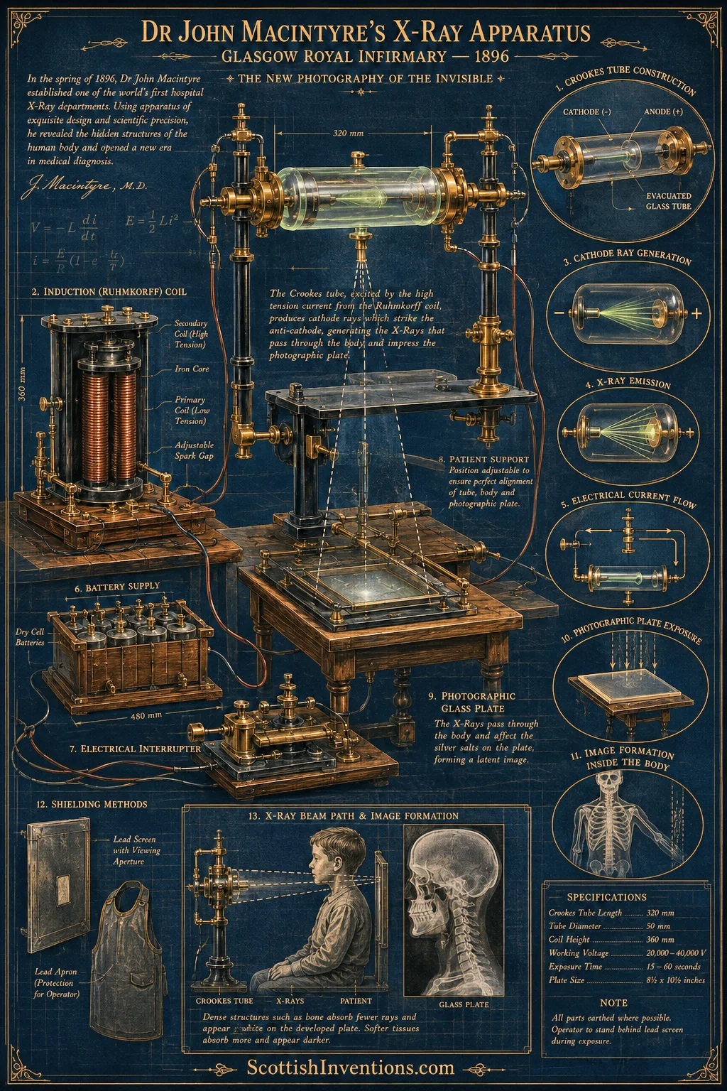
- Dry-cell batteries supplied the low-tension electrical current that powered the coil.
- A Ruhmkorff induction coil, with its primary and secondary windings around an iron core and an adjustable spark gap, stepped that current up to 20,000–40,000 volts.
- Those high-tension pulses were fed through an electrical interrupter into the Crookes tube — an evacuated glass vessel with a cathode and an anode, several hundred millimetres long.
- Inside the tube the discharge produced cathode rays that struck an anti-cathode, generating invisible X-rays which streamed out through the glass.
- The patient sat on a wooden support with a photographic glass plate beneath the body part being imaged. Exposures typically lasted 15 to 60 seconds.
- Dense structures such as bone absorbed more X-rays than soft tissue, leaving a lighter shadow on the developed silver-salt plate — the radiograph.
- Macintyre, unusually for the age, used lead screens and lead aprons to protect operators — a level of safety awareness that spared him the fate of many early radiologists commemorated on Hamburg's Radiation Martyrs' Monument.
The science of X-ray production had been enabled by two earlier Scottish contributions. James Clerk Maxwell's electromagnetic equations described the very radiation Röntgen was producing (Macintyre's own soft-tissue technique built directly on that theory — see our feature on James Clerk Maxwell and the first colour photograph). The wider culture of practical electrical medicine drew on the same tradition that gave Scotland the first electric light and the telephone.
"The Crookes tube, excited by the high-tension current from the Ruhmkorff coil, produces cathode rays which strike the anti-cathode, generating the X-Rays that pass through the body and impress the photographic plate."
Medical Breakthroughs
The Glasgow department was strikingly modern in the breadth of what it did. From its first years it offered plain-film radiography, radiotherapy (therapeutic X-rays), foreign-body localisation and stereoscopic radiography — the four pillars on which twentieth-century clinical radiology would be built. Macintyre published prolifically in 1896 alone; his papers appeared in the pioneering journal Archives of Clinical Skiagraphy, founded that year and later renamed the British Journal of Radiology.
Perhaps his most visually astonishing achievement was his X-ray cinematography. Working while the ink was still barely dry on Röntgen's paper, Macintyre produced X-ray moving pictures of the moving legs (specifically the knee joint) of a frog, and of a beating human heart, and — using bismuth as a contrast agent — of the movement of the digestive organs. Per the National Library of Scotland's Moving Image Archive (catalogue reference 0520), the film shows an "X-ray radiograph of adult, each picture taken in the 300th part of a second. A series of these pictures enable us to see a complete cycle of the movements of the heart." Filmed in 1896, the work was, in the archive's own words, "shown by Dr. John Macintyre at the London Royal Society 1897" — one of the earliest examples of moving-image radiography in history and a genuine rarity from the 1890s of film.
Macintyre was also unusually alert to the dangers of the new radiation. Where many early radiologists lost fingers, limbs and eventually their lives to X-ray burns and cancers, he introduced lead protection for himself and his staff — the reason he lived to old age when so many contemporaries did not.
World's Firsts
- First permanent hospital X-ray department — Glasgow Royal Infirmary, March 1896.
- First radiographic demonstration of a renal calculus verified at surgery — a landmark in diagnostic imaging.
- Foreign-body localisation — including a halfpenny lodged in a child's oesophagus, one of the earliest clinical uses of X-rays to guide treatment.
- Among the first radiographs of the spine, thorax, heart, lungs and kidney.
- One of the earliest X-ray cine films — a frog's leg, a beating human heart and the moving digestive organs, filmed 1896, shown at the Royal Society of London in 1897.
- Early stereoscopic radiography and therapeutic X-ray use inside a formal hospital department.
- Earlier still, in 1894–95, the first self-illuminated laryngeal endoscope.
Learn More: Interactive Panels
Tap any panel to expand.
+Learn more: Close: How the Crookes tube produced X-rays
A Crookes tube is a glass vessel from which almost all the air has been evacuated, with a metal cathode at one end and an anode at the other. When a very high voltage is applied across the electrodes, electrons stream from the cathode to the anode as a beam of "cathode rays". Where those cathode rays strike a metal target, they decelerate sharply and release part of their energy as X-rays. Röntgen's genius was to notice the effect; Macintyre's was to industrialise it into a routine hospital service.
+Learn more: Close: Why Glasgow, and not London or Vienna, was first
Several hospitals took clinical X-rays in early 1896 — John Hall-Edwards in Birmingham on 11 January, Dartmouth physicians in the United States on 3 February, teams in Vienna and Boston in March. What made Glasgow different was the combination of an existing hospital electrical department, mains power to every ward, and a Consulting Medical Electrician who was already both a working surgeon and a trained electrician. Vienna's famous Röntgen-Centrale only dates from 1898; its formal Central Radiology Institute from 1905. Massachusetts General Hospital's earliest images (mid-March 1896) were ad hoc work attached to medical photography, not a standing department. That is why Macintyre's March 1896 unit is widely credited as the world's first permanent hospital X-ray department.
+Learn more: Close: Röntgen and Macintyre exchanged tubes and letters
Röntgen and Macintyre were in direct contact. Röntgen reportedly asked (via Kelvin) how Macintyre had achieved his soft-tissue images, and later sent him one of his own X-ray tubes. Some of the early tubes used at Glasgow — donated by the widow of Dr Charles Read, a former GRI senior assistant radiologist — are preserved by the Royal College of Physicians and Surgeons of Glasgow.
+Learn more: Close: Radiography, fluoroscopy and radiotherapy — three uses, one tube
The same Crookes tube could be used three different ways. Radiography captured a still shadow on a photographic plate. Fluoroscopy let a clinician watch the moving shadow in real time on a fluorescent screen (the basis of the modern C-arm). Radiotherapy used the same X-rays deliberately to damage tissue — first attempted successfully on 24 November 1896 in Vienna by Leopold Freund on a five-year-old girl. All three modes were on offer in Macintyre's Glasgow department within a few years.
+Learn more: Close: The safety-conscious pioneer
Many early radiologists paid for the new technology with their lives. The Radiation Martyrs' Monument in Hamburg lists hundreds of doctors, nurses and technicians killed by X-ray induced burns, dermatitis and cancer in the first decades of the twentieth century. Macintyre — trained as an electrician before he became a physician — was unusually alert to the danger and introduced lead screens and lead aprons for himself and his staff from an early date, which is why he lived to 71.
+Learn more: Close: From plate to pixel — the modern imaging chain
Every step of the modern medical imaging chain has a direct descendant in Macintyre's Glasgow department. His glass plates evolved into film, then into computed radiography cassettes, then into today's flat-panel digital detectors. His Crookes tube evolved into the modern rotating-anode X-ray tube. His interpretation of shadow densities became the science of image reading; his radiotherapy became megavoltage linear-accelerator oncology; his fluoroscopy became the modern C-arm and the DSA suite; his cine radiography became CT, cardiac angiography and 4D imaging — with MRI, ultrasound and PET completing the family.
Timeline of the World's First Hospital X-Ray Department
2 October 1857
Born in Glasgow
John Macintyre is born in Glasgow's High Street, son of a Gaelic-speaking Fort William tailor and a mother said to be related to David Livingstone.
1878–1882
Medical training
After an early apprenticeship as an electrician, an aunt's bequest allows him to matriculate in medicine at the University of Glasgow. Graduates MB CM in 1882.
1885
Consulting Medical Electrician, GRI
Appointed to Glasgow Royal Infirmary in a role that uniquely combines his electrical and medical expertise.
1887
Department of Medical Electricity
Opens GRI's department for the application of medical electricity, wiring cables to every ward — one of the first hospitals with mains electricity throughout.
c.1894–95
Self-illuminated endoscope
Creates the first self-illuminated endoscope for examining the larynx, drawing directly on his combined interests in electric light and the throat.
8 November 1895
Röntgen discovers X-rays
In Würzburg, Wilhelm Röntgen discovers a mysterious new radiation and publishes his preliminary report at the end of December 1895.
January 1896
‘The New Light’
Macintyre lectures at Glasgow University on 'The New Light — X-rays', within weeks of Röntgen's paper reaching Britain via Lord Kelvin and his nephew J. T. Bottomley.
5 February 1896
First X-rays in Scotland
Macintyre, Bottomley and Lord Blythswood demonstrate X-rays and present 'On Röntgen X-Rays, or the new photography' to the Philosophical Society of Glasgow.
March 1896
World's first permanent hospital X-ray department
GRI managers consent to Macintyre establishing an X-ray unit within the electrical department — an ongoing radiographic service to patients, not a one-off experiment.
1896
Landmark clinical firsts
First radiograph of a renal calculus verified at surgery; radiograph of a halfpenny lodged in a child's oesophagus; among the first images of the spine, thorax, heart and kidney; and pioneering X-ray cine films of a frog's leg and moving human organs.
1897
Cine radiography at the Royal Society
Macintyre's X-ray moving pictures are shown at the Royal Society of London — one of the earliest examples of moving-image radiography.
1900–01
President of the Röntgen Society
Elected President of the Röntgen Society, the forerunner of the British Institute of Radiology.
1902
Purpose-built Electrical Pavilion
A new Electrical Pavilion opens at GRI as radiology rapidly expands; by 1903 the department is performing around 2,000 radiographs a year.
1920
Honoured by Glasgow
The University of Glasgow confers an LLD on Macintyre in recognition of his services to medicine and science.
29 October 1928
Death in Glasgow
Macintyre dies in Glasgow. His X-ray cine film — a genuine rarity from the 1890s — survives in the National Library of Scotland Moving Image Archive.
Today
Radiology everywhere
Every modern hospital in the world runs a radiology department descended from the model Macintyre established at Glasgow Royal Infirmary in 1896.
An Honest Claim
Distinguishing discovery from establishment
- Röntgen discovered X-rays on 8 November 1895 in Würzburg. Macintyre did not, and never claimed to.
- Glasgow was not first to take a clinical radiograph. That distinction belongs to John Hall-Edwards in Birmingham on 11 January 1896.
- Glasgow was the first permanent, organised hospital X-ray department — an ongoing service to patients rather than a one-off experiment. On that definition Macintyre's March 1896 unit is widely credited as, and quite possibly genuinely was, the world's first.
- The closest rivals are contemporaneous or later. Vienna's Röntgen-Centrale dates from 1898; its Central Radiology Institute from 1905. Boston's Massachusetts General Hospital produced its first images in mid-March 1896 as an ad hoc effort, not a formal department. Pennsylvania Hospital's organised X-ray provision dates to 1897.
Frequently Asked Questions
- Who was Dr John Macintyre?
- John Macintyre (1857–1928) was a Glasgow-born throat surgeon and pioneering medical electrician. Uniquely trained as both an apprentice electrician and a physician, he became Consulting Medical Electrician at Glasgow Royal Infirmary in 1885 and, in March 1896, founded what is widely credited as the world's first permanent hospital X-ray department.
- Did Scotland invent hospital radiology?
- Scotland did not discover X-rays — that credit belongs to Wilhelm Röntgen in Germany in November 1895. But Scotland can fairly claim to have invented hospital radiology as an organised clinical service. Macintyre's Glasgow Royal Infirmary department, founded in March 1896, is widely credited as the world's first permanent hospital X-ray department.
- What was the world's first hospital X-ray department?
- The X-ray department established by Dr John Macintyre at Glasgow Royal Infirmary in March 1896, within the hospital's existing electrical department. Unlike earlier one-off clinical radiographs elsewhere, it delivered an ongoing X-ray service to patients — the model every modern radiology department follows.
- Where was the first hospital X-ray department?
- Glasgow Royal Infirmary, Scotland. The department was housed within Macintyre's existing electrical department and later expanded into a purpose-built Electrical Pavilion, opened in 1902. By 1903 it was performing around 2,000 radiographs a year.
- What did Dr John Macintyre invent?
- Macintyre did not invent X-rays. His achievements were the world's first permanent hospital X-ray department; the first radiographic demonstration of a kidney stone (verified at surgery); early foreign-body localisation such as a halfpenny in a child's oesophagus; one of the earliest X-ray cine films (frog's leg and human heart, 1896); and, earlier, the first self-illuminated laryngeal endoscope (c.1894–95).
- How did early X-ray machines work?
- A Ruhmkorff induction coil, powered by dry-cell batteries, drove a high-voltage spark across a Crookes tube — an evacuated glass vessel with a cathode and anode. The electrical discharge produced invisible X-rays, which passed through the patient onto a photographic glass plate. Bones absorbed more X-rays than soft tissue, leaving a shadow image on the developed plate.
- When were X-rays first used in medicine?
- Within weeks of Röntgen's announcement in late December 1895. John Hall-Edwards took the first clinical radiograph in Birmingham on 11 January 1896; Dartmouth physicians took the first US clinical X-ray on 3 February 1896; and Macintyre and Bottomley demonstrated X-rays in Scotland on 5 February 1896. Macintyre's Glasgow department opened in March 1896.
- What happened at Glasgow Royal Infirmary in 1896?
- In January 1896 Macintyre lectured at Glasgow University on 'The New Light — X-rays'. On 5 February 1896 he, physicist James Thomson Bottomley and Lord Blythswood gave Scotland's first public X-ray demonstration. In March 1896 the GRI managers approved his proposal to open an X-ray unit, launching the world's first permanent hospital radiology department.
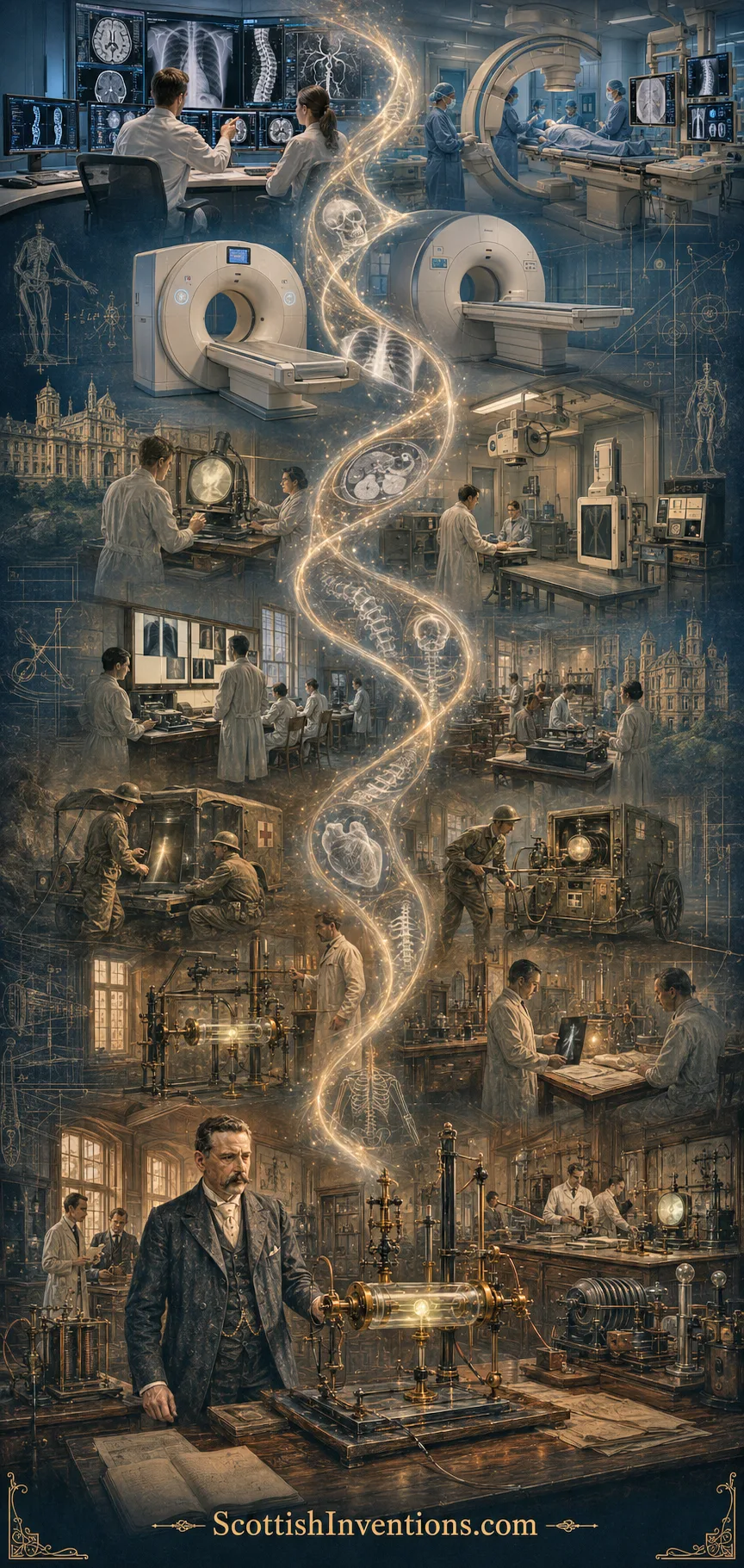
Lasting Legacy
The department Macintyre founded became a world leader in radiographic technique and grew into the modern radiology service at Glasgow Royal Infirmary. He was elected President of the Röntgen Society in 1900–01 — the forerunner of the British Institute of Radiology — awarded an LLD by the University of Glasgow in 1920 and was a Fellow of the Royal Society of Edinburgh. He died in Glasgow on 29 October 1928, still practising, still respected across two disciplines.
His wider legacy is enormous and, in a way, almost invisible — precisely because it has become the default. Every modern hospital in the world runs a radiology department. Every accident and emergency unit has an X-ray room. From plain film to computed tomography (CT), from fluoroscopy to angiography, from radiotherapy to interventional radiology, from the diagnostic ultrasound Ian Donald pioneered in the same city sixty years later to the MRI scanner built in Aberdeen in 1980, the entire structural template — a specialist hospital department housing imaging equipment, staffed by doctors, technologists and nurses, delivering a routine service to patients across the wards — is the one Macintyre invented in Glasgow in March 1896.
Macintyre's own X-ray cine film survives in the National Library of Scotland's Moving Image Archive (reference 0520). Early X-ray tubes he used are preserved by the Royal College of Physicians and Surgeons of Glasgow, and have been displayed in the RCPSG's exhibition "Our Science and Art: Visualising the Human Body". A 1970s documentary profile of Macintyre, narrated by John Scott — a past president of the Society of Radiographers who worked with him from 1914 to 1928 — was digitised in 1996 for the centenary of the department.
Why It Matters Today
More than a century on, medical imaging is one of the two or three great pillars of modern medicine, sitting alongside antisepsis, anaesthesia and pharmacology as a defining twentieth-century advance. Global radiology equipment is now a market measured in tens of billions of dollars; the average major hospital carries out hundreds of thousands of imaging examinations a year; and virtually no significant clinical decision in oncology, orthopaedics, neurology, cardiology or emergency medicine is now made without imaging.
All of that traces back to a simple institutional idea: that this new technology deserved its own department inside a working hospital, staffed by clinicians who knew both the physics and the patients. That idea is Macintyre's — and his city's. In an age when Scotland was already giving the world the telephone, the television, radar, the hypodermic syringe, chloroform anaesthesia, the cloud chamber, the kinetoscope and forensic fingerprinting, hospital radiology stands as one of the greatest Scottish contributions of all — because it changes the odds every time a patient walks into a hospital anywhere on earth.
"By 1903 the Electrical Pavilion was taking 2,000 radiographs each year and performing many fluoroscopic examinations."
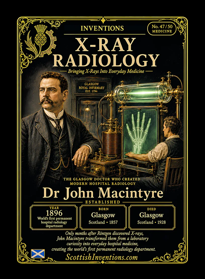
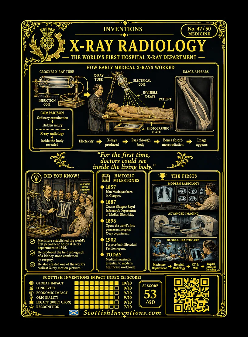
The X-Ray Radiology trading card
Card No. 47 of 50 in the Scottish Inventions Collection.
Continue Exploring
Scotland Changed Medicine Forever
From a single room in Glasgow Royal Infirmary, Dr John Macintyre transformed medicine by proving doctors could safely see inside the human body without surgery. Every modern X-ray, CT scan, fluoroscopy suite and radiology department traces its origins back to this remarkable Scottish breakthrough. Continue exploring the Scottish Inventions Collection and discover the remarkable ideas that changed the world.
Explore More Scottish InventionsSources
Tier 1 · Primary
- John F. Calder, The History of Radiology in Scotland, 1896–2000 (Dunedin Academic Press, 2001).
- Transactions of the Philosophical Society of Glasgow (1896) — "On Röntgen X-Rays, or the new photography".
- Archives of Clinical Skiagraphy (later British Journal of Radiology) — Macintyre's original 1896 papers.
Tier 2 · Scholarly and institutional
- A. M. K. Thomas & A. K. Banerjee, The History of Radiology (Oxford University Press, 2013); Adrian Thomas, "John Macintyre and the world's first x-ray department" (AuntMinnieEurope, 2013).
- Royal College of Physicians and Surgeons of Glasgow Heritage — "The World's First X-Ray Department" (2018).
- National Library of Scotland Moving Image Archive — "Dr Macintyre's X-Ray Film" (ref 0520).
- British Society for the History of Radiology and the British Institute of Radiology.
Tier 3 · General reference
- Wikipedia entries on Glasgow Royal Infirmary, Wilhelm Röntgen, John Hall-Edwards and the history of radiology.